Department of Ophthalmology, Shanghai Children's Hospital, School of Medicine, Shanghai Jiao Tong University, No.355 Luding Road, Shanghai, 200062, China.
Sci Rep. 2023 Feb 16;13(1):2780. doi: 10.1038/s41598-023-29816-1.
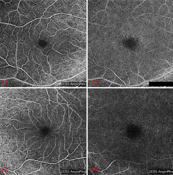
To compare and assess the choroidal and retinal microstructural vascularity in amblyopic eyes with the fellow eyes in anisometropic amblyopic children using angiography optical coherence tomography (Angio-OCT). Twenty-seven children (54 eyes; 5.59 ± 1.08 years old; 59.3% girls) were enrolled in this study. Choroidal thickness (CT) was measured with the use of the enhanced depth imaging mode in Angio-OCT. Parafoveal/peripapillary vascular density indices and the foveal avascular zone (FAZ) size were analyzed by MATLAB code programming on Angio-OCT images. The mean FAZ size of the amblyopic eyes were larger both in superficial and deep capillary plexus layer (SCPL/DCPL). Compared with the contralateral eyes (BCVA were normal), all the vascular density indices of SCPL and DCPL in the parafoveal and peripapillary zones were lower in the amblyopic eyes, however, the difference was insignificant (p > 0.05). No significant decrease was observed in four quadrants analyses of the amblyopic eyes (p > 0.05). Except for the measurement at 2000 µm and 1500 µm from the fovea in temple, CT in amblyopic eyes were significantly thicken than the fellow eyes (p < 0.05). Compared with the fellow eyes, the CT of certain areas were thicker in the amblyopic eyes. Though the FAZ size of the amblyopic eyes was larger in SCPL/DCPL layers, the retinal vascular density indices in SCPL/DCPL were lower in amblyopia eyes without statistical difference. Angio-OCT may be an effective way to evaluate the status of the choroidal and retinal vascular system in amblyopic children.
采用血管造影光学相干断层扫描(Angio-OCT)比较和评估屈光不正性弱视儿童弱视眼与对侧眼的脉络膜和视网膜微血管结构。本研究纳入 27 名儿童(54 只眼;5.59±1.08 岁;59.3%为女孩)。使用 Angio-OCT 的增强深度成像模式测量脉络膜厚度(CT)。使用 MATLAB 代码编程分析黄斑区及旁中心区血管密度指数和中心凹无血管区(FAZ)大小。与对侧眼(BCVA 正常)相比,弱视眼的浅层和深层毛细血管丛层(SCPL/DCPL)的平均 FAZ 面积均较大。与对侧眼相比,黄斑区及旁中心区 SCPL 和 DCPL 的所有血管密度指数均较低,但差异无统计学意义(p>0.05)。弱视眼四个象限的分析也没有明显的下降(p>0.05)。除了从颞侧距黄斑 2000 µm 和 1500 µm 处的测量值外,弱视眼的 CT 明显厚于对侧眼(p<0.05)。与对侧眼相比,弱视眼的某些区域的 CT 较厚。虽然 SCPL/DCPL 层的弱视眼 FAZ 面积较大,但 SCPL/DCPL 中的视网膜血管密度指数较低,差异无统计学意义。Angio-OCT 可能是评估弱视儿童脉络膜和视网膜血管系统状态的有效方法。