Department of Radiology, Klinikum Rechts Der Isar, School of Medicine, Technical University of Munich, Ismaninger Strasse 22, 81675, Munich, Germany.
Philips GmbH Market DACH, Hamburg, Germany.
Eur Radiol. 2023 Jul;33(7):4875-4884. doi: 10.1007/s00330-023-09472-9. Epub 2023 Feb 18.
To evaluate the diagnostic performance of an automated reconstruction algorithm combining MR imaging acquired using compressed SENSE (CS) with deep learning (DL) in order to reconstruct denoised high-quality images from undersampled MR images in patients with shoulder pain.
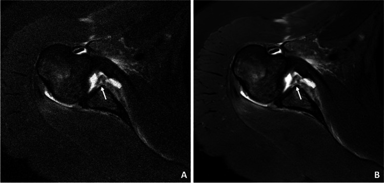
Prospectively, thirty-eight patients (14 women, mean age 40.0 ± 15.2 years) with shoulder pain underwent morphological MRI using a pseudo-random, density-weighted k-space scheme with an acceleration factor of 2.5 using CS only. An automated DL-based algorithm (CS DL) was used to create reconstructions of the same k-space data as used for CS reconstructions. Images were analyzed by two radiologists and assessed for pathologies, image quality, and visibility of anatomical landmarks using a 4-point Likert scale.
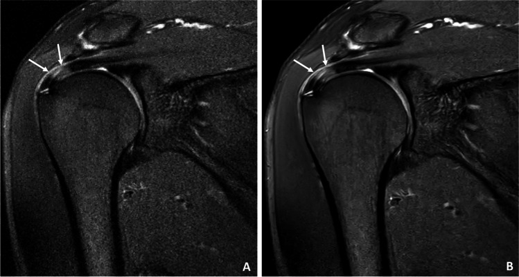
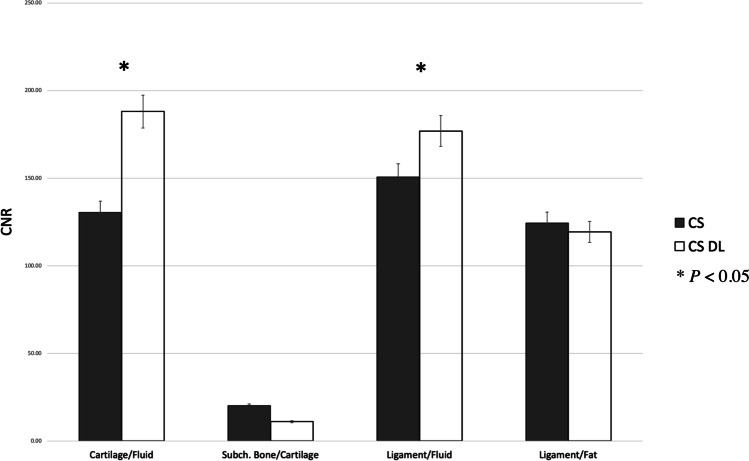
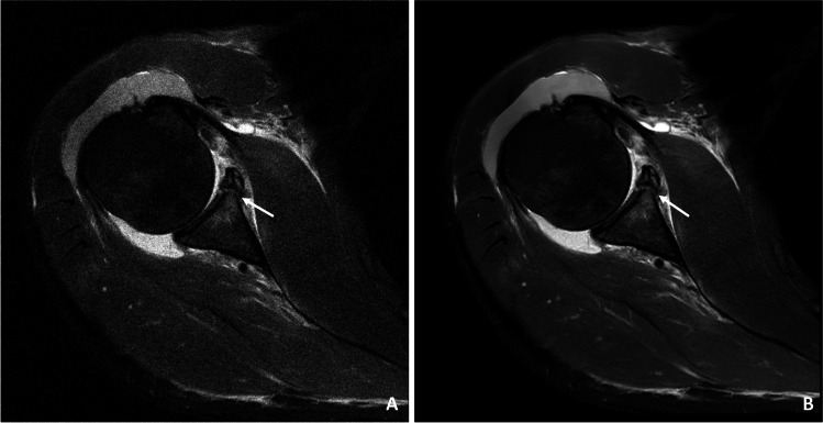
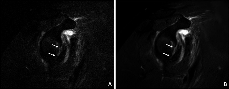
Overall agreement for the detection of pathologies between the CS DL reconstructions and CS images was substantial to almost perfect (κ 0.95 (95% confidence interval 0.82-1.00)). Image quality and the visibility of the rotator cuff, articular cartilage, and axillary recess were overall rated significantly higher for CS DL images compared to CS (p < 0.03). Contrast-to-noise ratios were significantly higher for cartilage/fluid (CS DL 198 ± 24.3, CS 130 ± 32.2, p = 0.02) and ligament/fluid (CS DL 184 ± 17.3, CS 141 ± 23.5, p = 0.03) and SNR values were significantly higher for ligaments and muscle of the CS DL reconstructions (p < 0.04).
Evaluation of shoulder pathologies was feasible using a DL-based algorithm for MRI reconstruction and denoising. In clinical routine, CS DL may be beneficial in particular for reducing image noise and may be useful for the detection and better discrimination of discrete pathologies. Assessment of shoulder pathologies was feasible with improved image quality as well as higher SNR using a compressed sensing deep learning-based framework for image reconstructions and denoising.
• Automated deep learning-based reconstructions showed a significant increase in signal-to-noise ratio and contrast-to-noise ratio (p < 0.04) with only a slight increase of reconstruction time of 40 s compared to CS. • All pathologies were accurately detected with no loss of diagnostic information or prolongation of the scan time. • Significant improvements of the image quality as well as the visibility of the rotator cuff, articular cartilage, and axillary recess were detected.
评估一种结合压缩感应(CS)与深度学习(DL)的自动重建算法在肩部疼痛患者中从欠采样磁共振成像重建去噪高质量图像的诊断性能。
前瞻性地,38 例肩部疼痛患者(14 名女性,平均年龄 40.0±15.2 岁)使用伪随机、密度加权的 k 空间方案进行形态学 MRI 检查,加速因子为 2.5,仅使用 CS。使用基于自动 DL 的算法(CS DL)来创建与 CS 重建相同的 k 空间数据的重建。由两名放射科医生对图像进行分析,并使用 4 分制量表评估病变、图像质量和解剖标志的可见度。
CS DL 重建图像与 CS 图像之间对病变的检测总体一致性为高度一致至几乎完美(κ 0.95(95%置信区间 0.82-1.00))。与 CS 相比,CS DL 图像的整体图像质量和肩袖、关节软骨以及腋窝的可视性评分明显更高(p<0.03)。软骨/液体(CS DL 198±24.3,CS 130±32.2,p=0.02)和韧带/液体(CS DL 184±17.3,CS 141±23.5,p=0.03)的对比噪声比明显更高,并且 CS DL 重建的韧带和肌肉的 SNR 值明显更高(p<0.04)。
使用基于 DL 的算法进行 MRI 重建和去噪可以评估肩部病变。在临床常规中,CS DL 可能特别有益于降低图像噪声,并且可能有助于离散病变的检测和更好的区分。使用压缩感知深度学习框架进行图像重建和去噪,可以改善图像质量和提高信噪比,从而实现肩部病变的评估。
与 CS 相比,基于自动深度学习的重建显示出 SNR 和对比噪声比的显著增加(p<0.04),而重建时间仅增加了 40 秒。
所有病变均准确检测,无诊断信息丢失或扫描时间延长。
检测到图像质量以及肩袖、关节软骨和腋窝的可见度的显著改善。