Ceron Hartmann Giovani, Costa Brancalhão Rose Meire, Chasko Ribeiro Lucinéia de Fátima, Carrinho Ayroza Rangel Ana Lúcia, Giampietro-Brandão Christian
Department of Biological Sciences and Health of Western Paraná State University (UNIOESTE), Brazil.
Iran Endod J. 2019 Summer;14(3):190-196. doi: 10.22037/iej.v14i3.22690.
This histological study analyzed silk sericin as a potential direct pulp capping biomaterial in contact with pulp and comparing its response to calcium hydroxide.
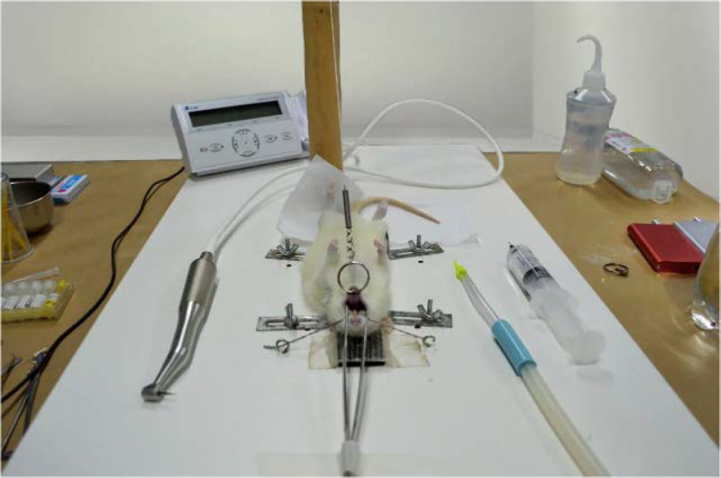
Twenty maxillary first molars from Wistar male rats were used, with 60 days of age, between 200 and 300 gr, which were divided in 4 groups (=5): G1 and G3, controls, capped with calcium hydroxide in 7 and 30 days, respectively; G2 and G4, capped with silk sericin in 7 and 30 days, respectively. Circular cavities were prepared for pulp exposure, where capping materials were applied, being posteriorly restored with glass ionomer cement. After completion of each observation period, the animals were sacrificed and molars were histologically processed for analysis in light microscopy to evaluate presence of necrosis in pulp tissue, inflammatory cells infiltration and tertiary dentin formation. Data analysis was carried out using Kruskal-Wallis and Dunn's post hoc tests.
After 7 days, there was less necrosis and inflammatory cells infiltration in G1 when compared to G2 (=0.007 and =0.008, respectively). After 30 days, a sample of G3 induced tertiary dentin formation and G4 showed decrease in inflammation (=0.041) compared to G2.
Among the determined experiment conditions, it was concluded that contact between silk sericin and pulp tissue showed improved inflammatory response throughout treatment and new cells proliferation. However, silk sericin adhibition in pure form did not show capability for induction of tertiary dentin formation.
本组织学研究分析了丝胶蛋白作为一种潜在的直接盖髓生物材料与牙髓接触的情况,并将其反应与氢氧化钙进行比较。
使用20颗来自60日龄、体重在200至300克之间的雄性Wistar大鼠的上颌第一磨牙,将其分为4组(每组5颗):G1和G3为对照组,分别在7天和30天后用氢氧化钙覆盖;G2和G4分别在7天和30天后用丝胶蛋白覆盖。制备圆形洞以暴露牙髓,在洞中应用盖髓材料,随后用玻璃离子水门汀进行修复。在每个观察期结束后,处死动物,对磨牙进行组织学处理,以便在光学显微镜下分析,以评估牙髓组织中的坏死情况、炎症细胞浸润和第三期牙本质形成。使用Kruskal-Wallis和Dunn事后检验进行数据分析。
7天后,与G2相比,G1中的坏死和炎症细胞浸润较少(分别为P = 0.007和P = 0.008)。30天后,G3的一个样本诱导了第三期牙本质形成,与G2相比,G4的炎症有所减轻(P = 0.041)。
在确定的实验条件下,得出的结论是,丝胶蛋白与牙髓组织的接触在整个治疗过程中显示出改善的炎症反应和新细胞增殖。然而,纯形式的丝胶蛋白吸附并未显示出诱导第三期牙本质形成的能力。