Mortberg Meredith A, Gentile Juliana E, Nadaf Naeem, Vanderburg Charles, Simmons Sean, Dubinsky Dan, Slamin Adam, Maldonado Salome, Petersen Caroline L, Jones Nichole, Kordasiewicz Holly B, Zhao Hien T, Vallabh Sonia M, Minikel Eric Vallabh
Stanley Center for Psychiatric Research, Broad Institute of MIT and Harvard,Cambridge, MA, 02142, USA.
Genomics Platform, Broad Institute of MIT and Harvard, Cambridge, MA, 02141, USA.
bioRxiv. 2023 Feb 14:2023.02.14.528473. doi: 10.1101/2023.02.14.528473.
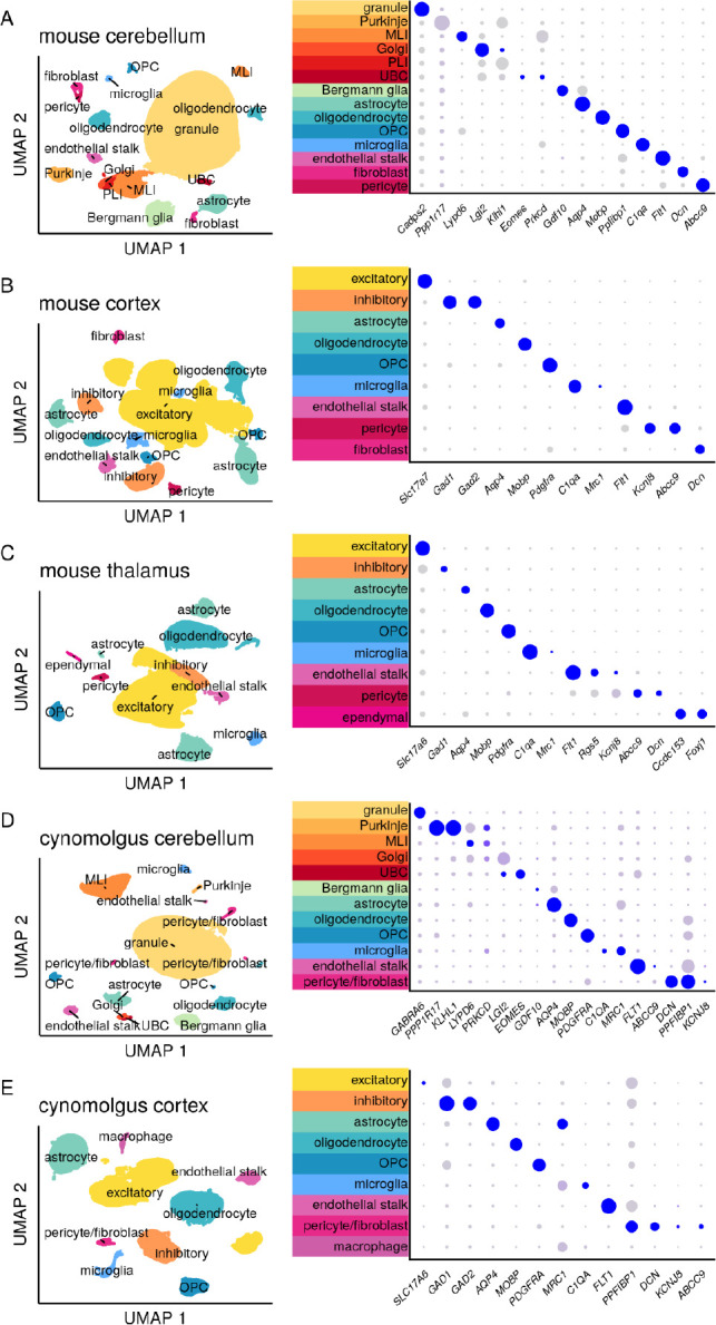
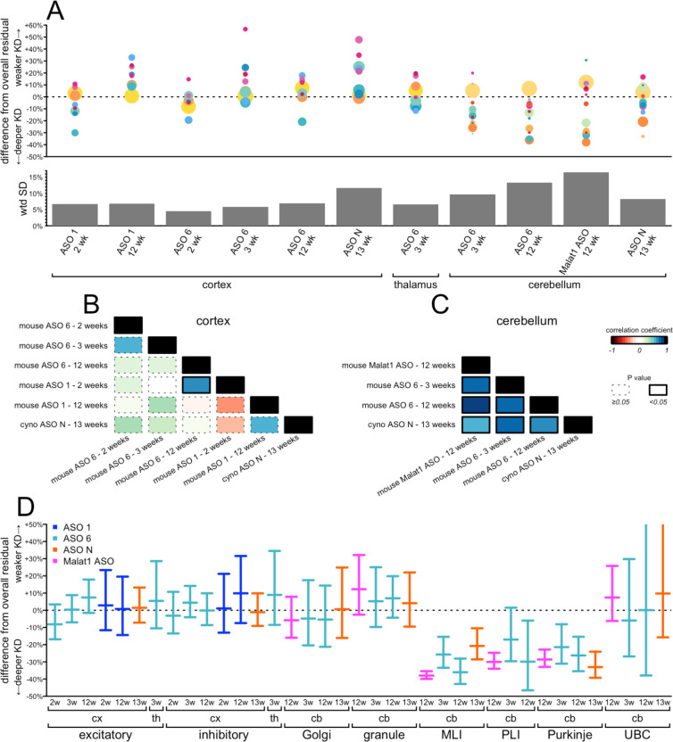
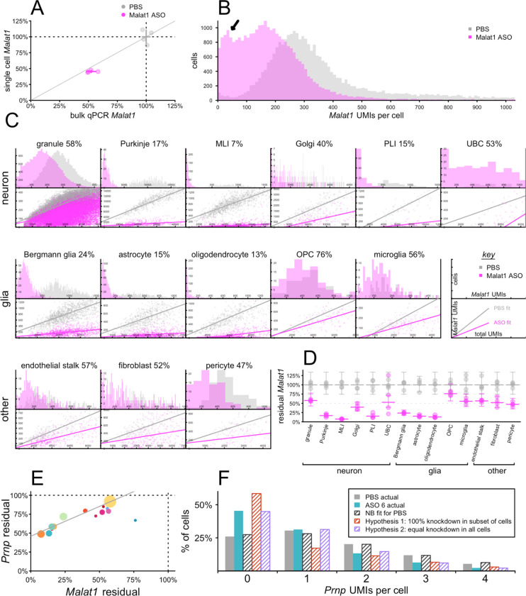
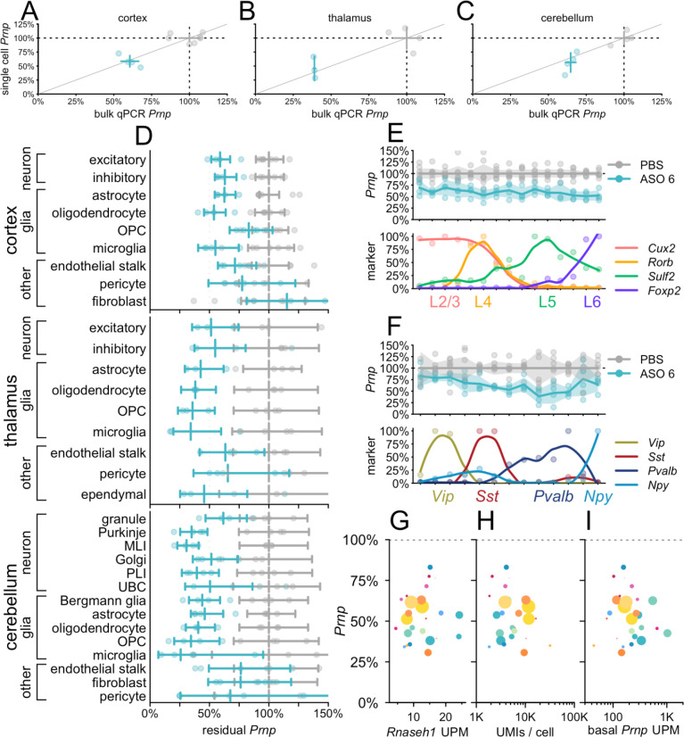
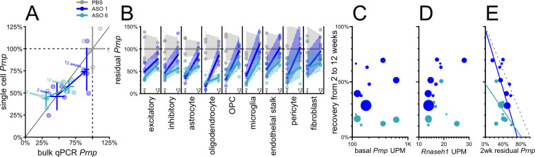
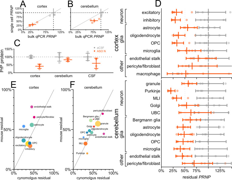
Antisense oligonucleotides (ASOs) dosed into cerebrospinal fluid (CSF) distribute broadly throughout the brain and hold the promise of treating myriad brain diseases by modulating RNA. CNS tissue is not routinely biopsied in living individuals, leading to reliance on CSF biomarkers to inform on drug target engagement. Animal models can link CSF biomarkers to brain parenchyma, but our understanding of how individual cells contribute to bulk tissue signal is limited. Here we employed single nucleus transcriptomics on tissue from mice treated with RNase H1 ASOs against and and macaques treated with an ASO against . Activity was observed in every cell type, though sometimes with substantial differences in magnitude. Single cell RNA count distributions implied target suppression in every single sequenced cell, rather than intense knockdown in only some cells. Duration of action up to 12 weeks post-dose differed across cell types, being shorter in microglia than in neurons. Suppression in neurons was generally similar to, or more robust than, the bulk tissue. In macaques, PrP in CSF was lowered 40% in conjunction with knockdown across all cell types including neurons, arguing that a CSF biomarker readout is likely to reflect disease-relevant cells in a neuronal disorder.
注入脑脊液(CSF)中的反义寡核苷酸(ASO)可广泛分布于整个大脑,并有望通过调节RNA来治疗多种脑部疾病。在活体个体中,中枢神经系统(CNS)组织通常不会进行活检,这导致人们依赖脑脊液生物标志物来了解药物靶点的作用情况。动物模型可以将脑脊液生物标志物与脑实质联系起来,但我们对单个细胞如何对整体组织信号产生影响的了解有限。在这里,我们对用针对 和 的核糖核酸酶H1 ASO处理的小鼠以及用针对 的ASO处理的猕猴的组织进行了单核转录组学分析。在每种细胞类型中都观察到了活性,尽管有时在程度上存在显著差异。单细胞RNA计数分布表明,在每个测序细胞中都存在靶点抑制,而不是仅在某些细胞中出现强烈的敲低。给药后长达12周的作用持续时间在不同细胞类型中有所不同,小胶质细胞中的持续时间比神经元中的短。神经元中的抑制作用通常与整体组织相似,或比整体组织更强。在猕猴中,脑脊液中的PrP降低了40%,同时在包括神经元在内的所有细胞类型中 均被敲低,这表明脑脊液生物标志物读数可能反映了神经疾病中与疾病相关的细胞。