Department of Mechanical Engineering, Virginia Tech, Blacksburg, VA 24061.
School of Mathematical and Computational Sciences, Indian Association for the Cultivation of Science, Jadavpur, Kolkata 700032, India.
Proc Natl Acad Sci U S A. 2023 Mar 7;120(10):e2120536120. doi: 10.1073/pnas.2120536120. Epub 2023 Feb 27.
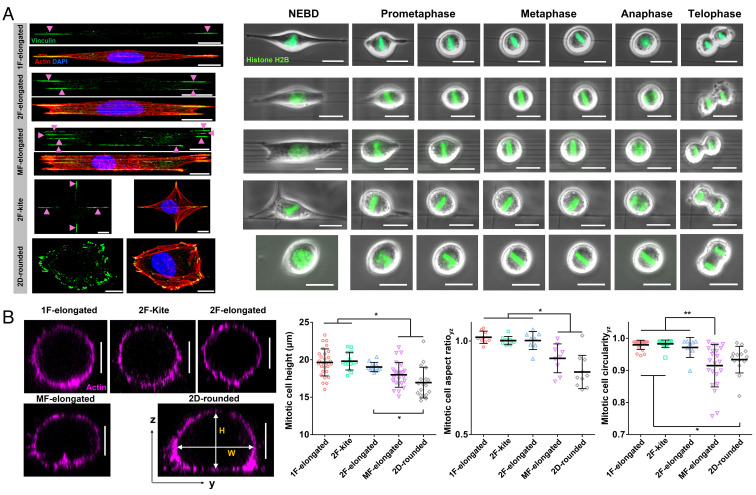
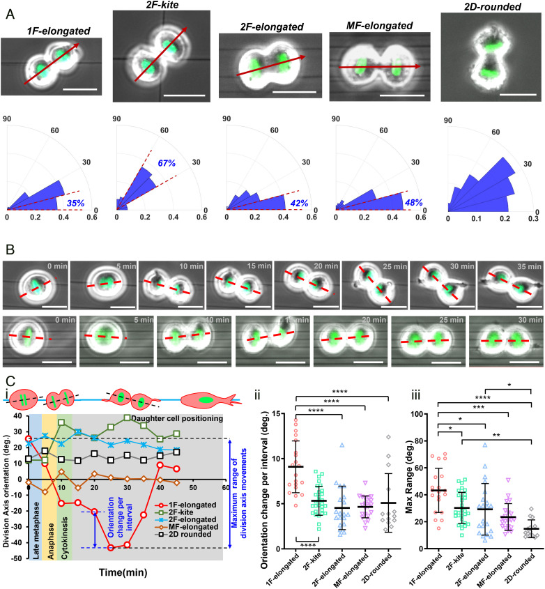
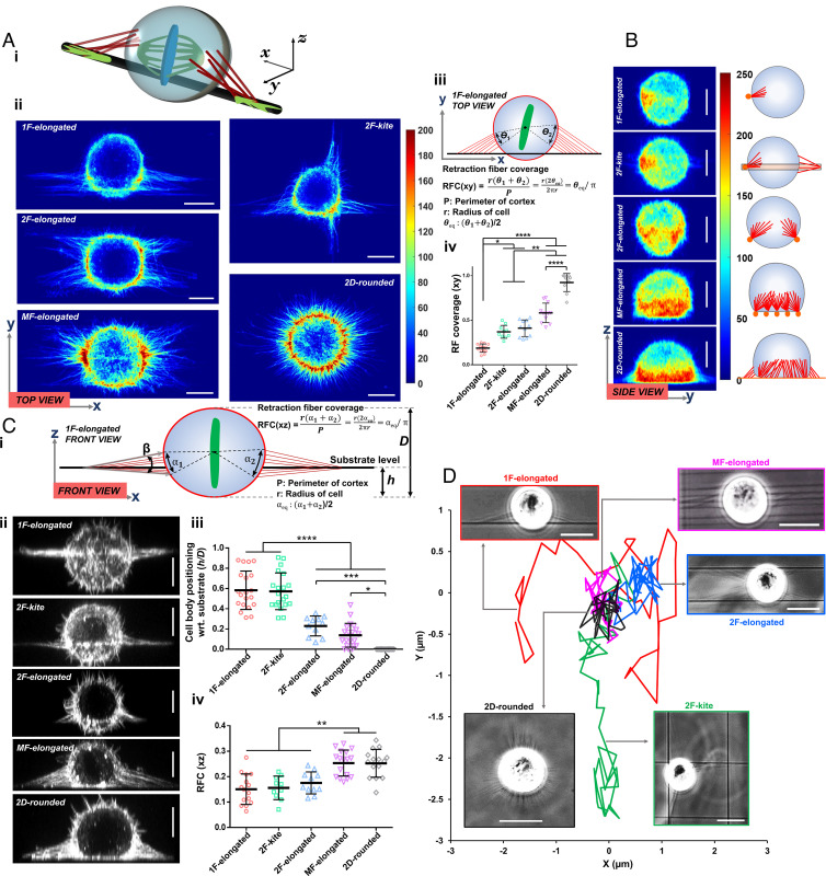
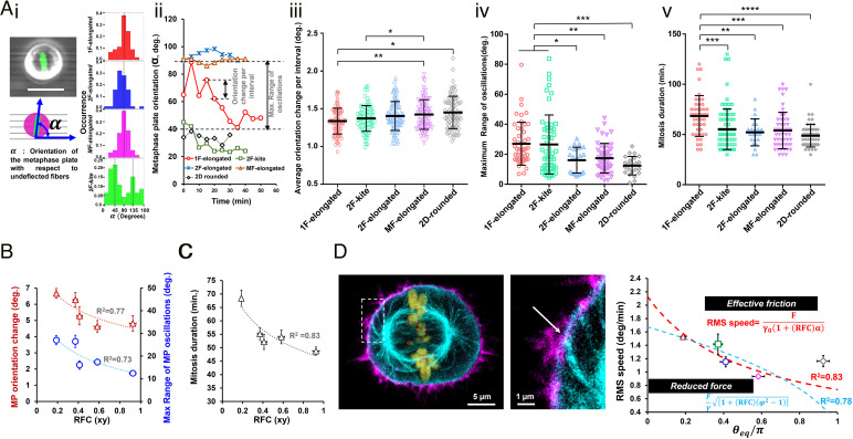
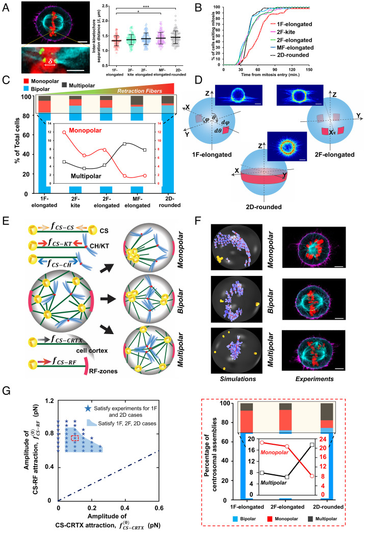
During mitosis, cells round up and utilize the interphase adhesion sites within the fibrous extracellular matrix (ECM) as guidance cues to orient the mitotic spindles. Here, using suspended ECM-mimicking nanofiber networks, we explore mitotic outcomes and error distribution for various interphase cell shapes. Elongated cells attached to single fibers through two focal adhesion clusters (FACs) at their extremities result in perfect spherical mitotic cell bodies that undergo significant 3-dimensional (3D) displacement while being held by retraction fibers (RFs). Increasing the number of parallel fibers increases FACs and retraction fiber-driven stability, leading to reduced 3D cell body movement, metaphase plate rotations, increased interkinetochore distances, and significantly faster division times. Interestingly, interphase kite shapes on a crosshatch pattern of four fibers undergo mitosis resembling single-fiber outcomes due to rounded bodies being primarily held in position by RFs from two perpendicular suspended fibers. We develop a cortex-astral microtubule analytical model to capture the retraction fiber dependence of the metaphase plate rotations. We observe that reduced orientational stability, on single fibers, results in increased monopolar mitotic defects, while multipolar defects become dominant as the number of adhered fibers increases. We use a stochastic Monte Carlo simulation of centrosome, chromosome, and membrane interactions to explain the relationship between the observed propensity of monopolar and multipolar defects and the geometry of RFs. Overall, we establish that while bipolar mitosis is robust in fibrous environments, the nature of division errors in fibrous microenvironments is governed by interphase cell shapes and adhesion geometries.
在有丝分裂过程中,细胞变圆,并利用细胞间的黏附位点作为导向线索,使有丝分裂纺锤体定向排列。在这里,我们使用悬浮的类似细胞外基质 (ECM) 的纳米纤维网络,探索了各种细胞间形状的有丝分裂结果和错误分布。细长的细胞通过其两端的两个焦点黏附簇 (FAC) 附着在单根纤维上,导致有丝分裂细胞体呈完美的球形,当被牵拉纤维 (RF) 牵拉时,会发生显著的三维 (3D) 位移。增加平行纤维的数量会增加 FAC 和牵拉纤维驱动的稳定性,从而减少 3D 细胞体的运动、中期板的旋转、动粒间距离的增加以及分裂时间的显著加快。有趣的是,在四条纤维的交叉模式上呈风筝形状的细胞间期经历有丝分裂,类似于单纤维的结果,因为圆形的细胞体主要通过来自两个垂直悬挂纤维的牵拉纤维保持在适当的位置。我们开发了一个皮质-星体微管分析模型来捕捉中期板旋转的牵拉纤维依赖性。我们观察到,在单纤维上,定向稳定性降低会导致单极有丝分裂缺陷增加,而随着附着纤维数量的增加,多极缺陷变得占主导地位。我们使用中心体、染色体和膜相互作用的随机蒙特卡罗模拟来解释观察到的单极和多极缺陷的倾向与牵拉纤维几何形状之间的关系。总的来说,我们建立了这样的观点,即虽然在纤维环境中双极有丝分裂是稳健的,但纤维微环境中分裂错误的性质是由细胞间期的形状和黏附几何形状决定的。