• 文献检索
  • 文档翻译
  • 深度研究
  • 学术资讯
  • Suppr Zotero 插件Zotero 插件
  • 邀请有礼
  • 套餐&价格
  • 历史记录
应用&插件
Suppr Zotero 插件Zotero 插件浏览器插件Mac 客户端Windows 客户端微信小程序
定价
高级版会员购买积分包购买API积分包
服务
文献检索文档翻译深度研究API 文档MCP 服务
关于我们
关于 Suppr公司介绍联系我们用户协议隐私条款
关注我们

Suppr 超能文献

核心技术专利:CN118964589B侵权必究
粤ICP备2023148730 号-1Suppr @ 2026

文献检索

告别复杂PubMed语法,用中文像聊天一样搜索,搜遍4000万医学文献。AI智能推荐,让科研检索更轻松。

立即免费搜索

文件翻译

保留排版,准确专业,支持PDF/Word/PPT等文件格式,支持 12+语言互译。

免费翻译文档

深度研究

AI帮你快速写综述,25分钟生成高质量综述,智能提取关键信息,辅助科研写作。

立即免费体验

CAMSAP3 对扩展微管晶格的偏好有助于稳定微管的负端。

Preference of CAMSAP3 for expanded microtubule lattice contributes to stabilization of the minus end.

机构信息

Department of Biological Sciences, Graduate School of Science, The University of Tokyo, Tokyo, Japan.

Department of Biological Sciences, Graduate School of Science, The University of Tokyo, Tokyo, Japan

出版信息

Life Sci Alliance. 2023 Mar 9;6(5). doi: 10.26508/lsa.202201714. Print 2023 May.

DOI:10.26508/lsa.202201714
PMID:36894175
原文链接:https://pmc.ncbi.nlm.nih.gov/articles/PMC9998277/
Abstract

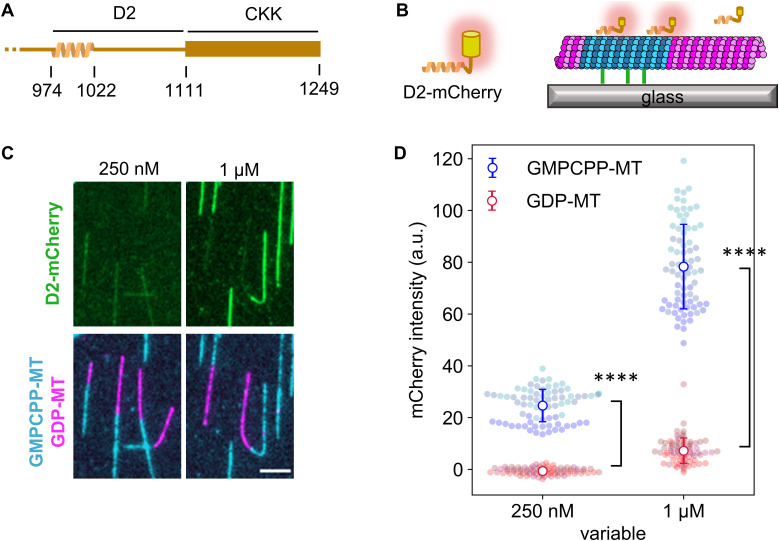
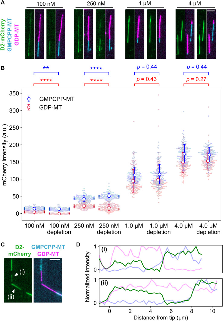
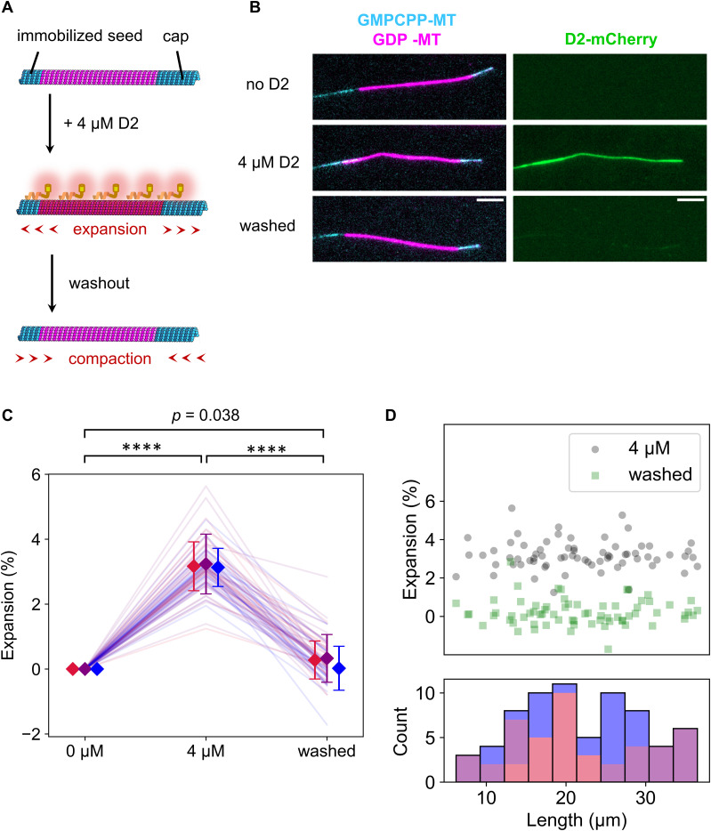
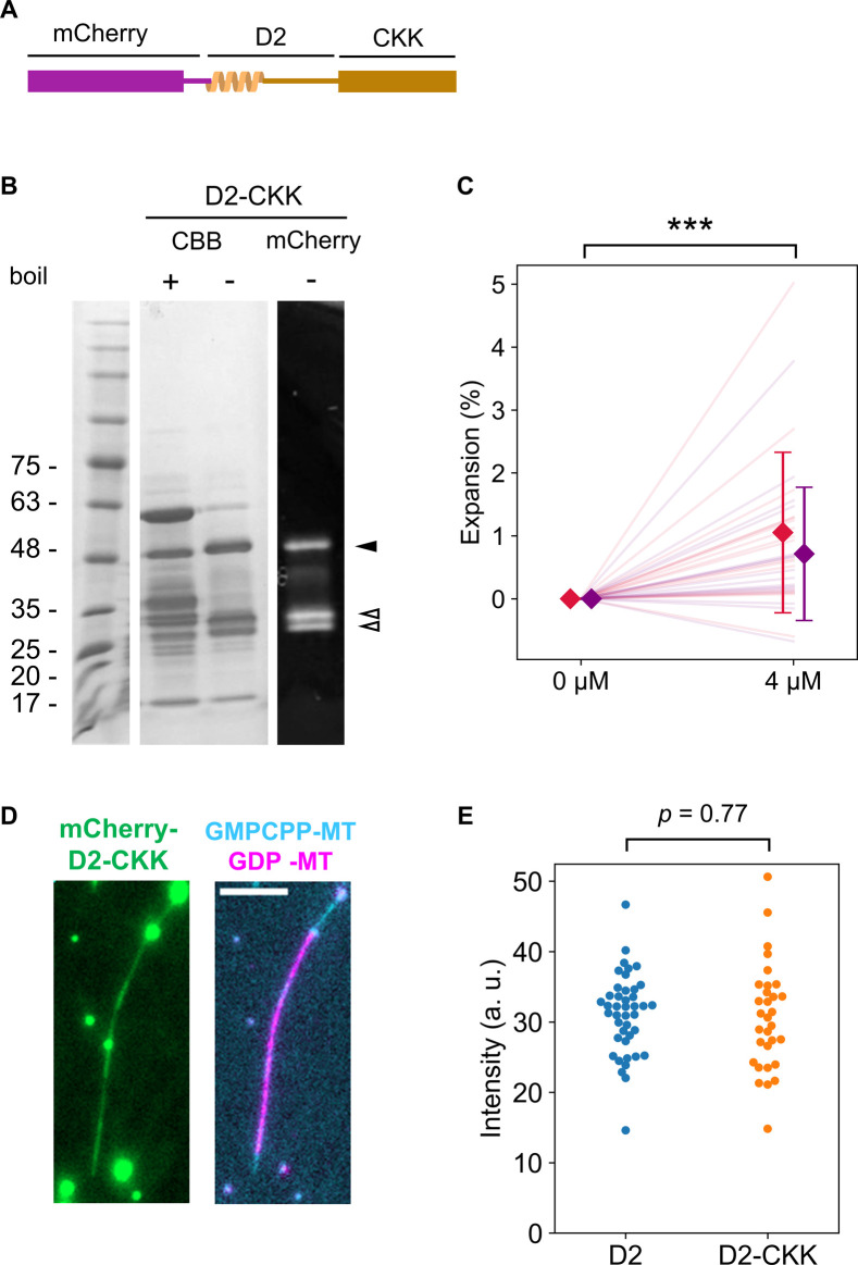
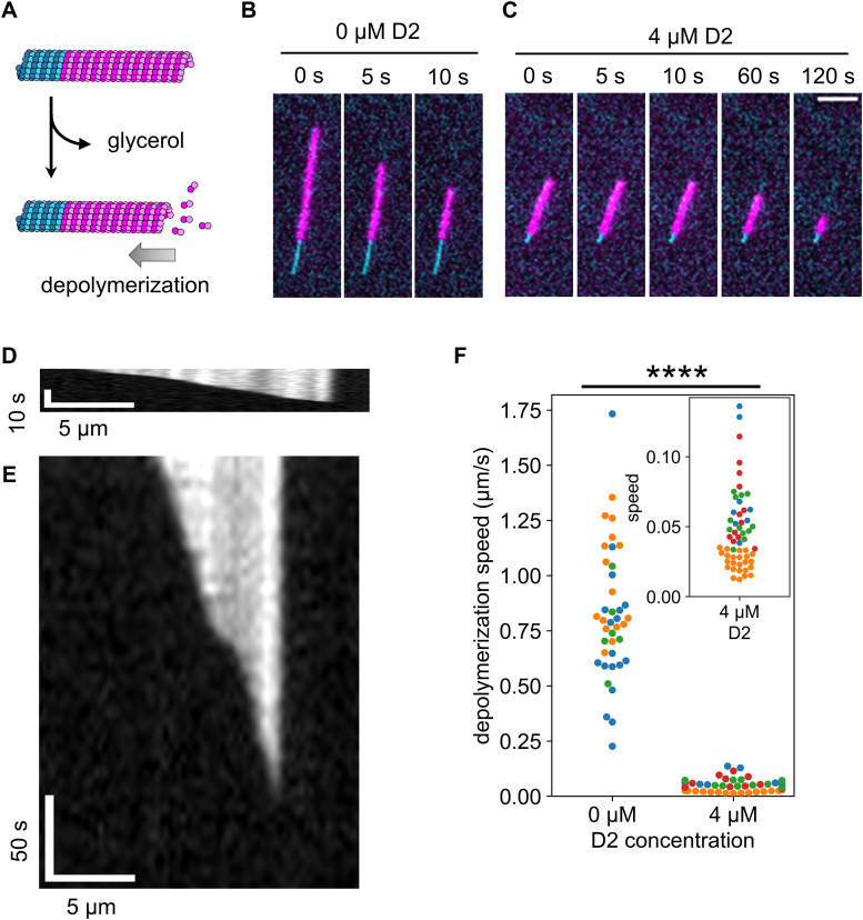
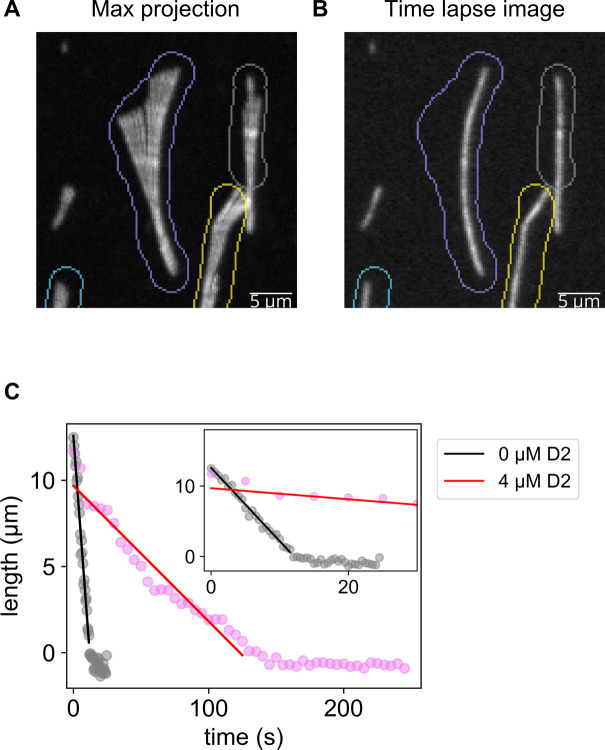
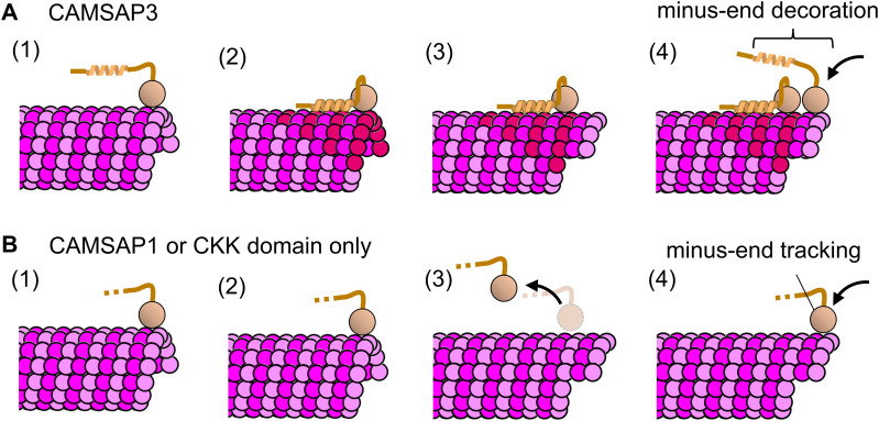
CAMSAPs are proteins that show microtubule minus-end-specific localization, decoration, and stabilization. Although the mechanism for minus-end recognition via their C-terminal CKK domain has been well described in recent studies, it is unclear how CAMSAPs stabilize microtubules. Our several binding assays revealed that the D2 region of CAMSAP3 specifically binds to microtubules with the expanded lattice. To investigate the relationship between this preference and the stabilization effect of CAMSAP3, we precisely measured individual microtubule lengths and found that D2 binding expanded the microtubule lattice by ∼3%. Consistent with the notion that the expanded lattice is a common feature of stable microtubules, the presence of D2 slowed the microtubule depolymerization rate to ∼1/20, suggesting that the D2-triggered lattice expansion stabilizes microtubules. Combining these results, we propose that CAMSAP3 stabilizes microtubules by lattice expansion upon D2 binding, which further accelerates the recruitment of other CAMSAP3 molecules. Because only CAMSAP3 has D2 and the highest microtubule-stabilizing effect among mammalian CAMSAPs, our model also explains the molecular basis for the functional diversity of CAMSAP family members.

摘要

CAMSAPs 是一类具有微管负端特异性定位、修饰和稳定作用的蛋白。虽然其 C 端 CKK 结构域通过负端识别的机制在最近的研究中已被很好地描述,但 CAMSAPs 如何稳定微管尚不清楚。我们的几项结合实验表明,CAMSAP3 的 D2 区特异性结合具有扩展晶格的微管。为了研究这种偏好与 CAMSAP3 稳定作用之间的关系,我们精确测量了单个微管的长度,发现 D2 结合将微管晶格扩展了约 3%。与扩展晶格是稳定微管的共同特征的观点一致,D2 的存在将微管解聚速率降低到约 1/20,表明 D2 触发的晶格扩展稳定了微管。综合这些结果,我们提出 CAMSAP3 通过 D2 结合引发晶格扩展来稳定微管,这进一步加速了其他 CAMSAP3 分子的募集。由于只有 CAMSAP3 具有 D2 结构域和在哺乳动物 CAMSAPs 中具有最高的微管稳定作用,我们的模型还解释了 CAMSAP 家族成员功能多样性的分子基础。

https://cdn.ncbi.nlm.nih.gov/pmc/blobs/7230/9998277/bafa67634829/LSA-2022-01714_FigS5.jpg
https://cdn.ncbi.nlm.nih.gov/pmc/blobs/7230/9998277/206f85e207e6/LSA-2022-01714_FigS1.jpg
https://cdn.ncbi.nlm.nih.gov/pmc/blobs/7230/9998277/89369db87cb3/LSA-2022-01714_Fig1.jpg
https://cdn.ncbi.nlm.nih.gov/pmc/blobs/7230/9998277/f46109daf462/LSA-2022-01714_Fig2.jpg
https://cdn.ncbi.nlm.nih.gov/pmc/blobs/7230/9998277/4e08256a2ba8/LSA-2022-01714_Fig3.jpg
https://cdn.ncbi.nlm.nih.gov/pmc/blobs/7230/9998277/26001eda2671/LSA-2022-01714_Fig4.jpg
https://cdn.ncbi.nlm.nih.gov/pmc/blobs/7230/9998277/b92b7cce6e8f/LSA-2022-01714_FigS2.jpg
https://cdn.ncbi.nlm.nih.gov/pmc/blobs/7230/9998277/9ad3aa4fa6f4/LSA-2022-01714_Fig5.jpg
https://cdn.ncbi.nlm.nih.gov/pmc/blobs/7230/9998277/cae6c6a6ee08/LSA-2022-01714_FigS3.jpg
https://cdn.ncbi.nlm.nih.gov/pmc/blobs/7230/9998277/03a26af8be49/LSA-2022-01714_FigS4.jpg
https://cdn.ncbi.nlm.nih.gov/pmc/blobs/7230/9998277/e50647f35bec/LSA-2022-01714_Fig6.jpg
https://cdn.ncbi.nlm.nih.gov/pmc/blobs/7230/9998277/bafa67634829/LSA-2022-01714_FigS5.jpg

相似文献

1
Preference of CAMSAP3 for expanded microtubule lattice contributes to stabilization of the minus end.CAMSAP3 对扩展微管晶格的偏好有助于稳定微管的负端。
Life Sci Alliance. 2023 Mar 9;6(5). doi: 10.26508/lsa.202201714. Print 2023 May.
2
A structural model for microtubule minus-end recognition and protection by CAMSAP proteins.CAMSAP蛋白对微管负端识别与保护的结构模型。
Nat Struct Mol Biol. 2017 Nov;24(11):931-943. doi: 10.1038/nsmb.3483. Epub 2017 Oct 9.
3
Structural Basis of Formation of the Microtubule Minus-End-Regulating CAMSAP-Katanin Complex.微管负端调节 CAMSAP-卡坦in 复合物形成的结构基础。
Structure. 2018 Mar 6;26(3):375-382.e4. doi: 10.1016/j.str.2017.12.017. Epub 2018 Jan 26.
4
Regulation of microtubule minus-end dynamics by CAMSAPs and Patronin.CAMSAPs 和 Patronin 对微管负端动力学的调节。
Proc Natl Acad Sci U S A. 2014 Apr 22;111(16):5860-5. doi: 10.1073/pnas.1404133111. Epub 2014 Mar 26.
5
Microtubule minus-end stabilization by polymerization-driven CAMSAP deposition.微管负端通过聚合驱动的 CAMSAP 沉积稳定化。
Dev Cell. 2014 Feb 10;28(3):295-309. doi: 10.1016/j.devcel.2014.01.001. Epub 2014 Jan 30.
6
CAMSAPs and nucleation-promoting factors control microtubule release from γ-TuRC.CAMSAPs 和成核促进因子控制 γ-TuRC 从小管释放。
Nat Cell Biol. 2024 Mar;26(3):404-420. doi: 10.1038/s41556-024-01366-2. Epub 2024 Feb 29.
7
CAMSAP3 orients the apical-to-basal polarity of microtubule arrays in epithelial cells.CAMSAP3确定上皮细胞中微管阵列从顶端到基部的极性。
Proc Natl Acad Sci U S A. 2016 Jan 12;113(2):332-7. doi: 10.1073/pnas.1520638113. Epub 2015 Dec 29.
8
Control of apico-basal epithelial polarity by the microtubule minus-end-binding protein CAMSAP3 and spectraplakin ACF7.微管负端结合蛋白CAMSAP3和光谱斑蛋白ACF7对上皮细胞顶-基极性的调控
J Cell Sci. 2016 Nov 15;129(22):4278-4288. doi: 10.1242/jcs.194878. Epub 2016 Oct 17.
9
CAMSAP2 and CAMSAP3 localize at microtubule intersections to regulate the spatial distribution of microtubules.CAMSAP2 和 CAMSAP3 定位于微管交点,以调节微管的空间分布。
J Mol Cell Biol. 2024 Jan 17;15(8). doi: 10.1093/jmcb/mjad050.
10
Cadherin 23-C Regulates Microtubule Networks by Modifying CAMSAP3's Function.钙黏蛋白 23-C 通过调节 CAMSAP3 的功能来调节微管网络。
Sci Rep. 2016 Jun 28;6:28706. doi: 10.1038/srep28706.

引用本文的文献

1
MATCAP1 preferentially binds an expanded tubulin conformation to generate detyrosinated and ΔC2 α-tubulin.MATCAP1优先结合扩展的微管蛋白构象以生成去酪氨酸化和ΔC2α-微管蛋白。
bioRxiv. 2025 Aug 18:2025.08.14.670257. doi: 10.1101/2025.08.14.670257.
2
Diverse microtubule-binding repeats regulate TPX2 activities at distinct locations within the spindle.多种微管结合重复序列在纺锤体内不同位置调节TPX2活性。
J Cell Biol. 2025 Mar 3;224(3). doi: 10.1083/jcb.202404025. Epub 2025 Jan 16.
3
CAMSAP2 is required for bridging fiber assembly to ensure mitotic spindle assembly and chromosome segregation in human epithelial Caco-2 cells.

本文引用的文献

1
CAMSAP2 organizes a γ-tubulin-independent microtubule nucleation centre through phase separation.CAMSAP2 通过相分离组织一个γ-微管蛋白独立的微管核中心。
Elife. 2022 Jun 28;11:e77365. doi: 10.7554/eLife.77365.
2
A hydrodynamic instability drives protein droplet formation on microtubules to nucleate branches.一种流体动力学不稳定性驱动微管上蛋白质液滴形成以产生分支。
Nat Phys. 2021 Apr;17(4):493-498. doi: 10.1038/s41567-020-01141-8. Epub 2021 Jan 28.
3
Structural transitions in the GTP cap visualized by cryo-electron microscopy of catalytically inactive microtubules.
在人上皮Caco-2细胞中,桥连纤维组装需要CAMSAP2以确保有丝分裂纺锤体组装和染色体分离。
PLoS One. 2025 Jan 9;20(1):e0308150. doi: 10.1371/journal.pone.0308150. eCollection 2025.
4
Structure of blood cell-specific tubulin and demonstration of dimer spacing compaction in a single protofilament.血细胞特异性微管蛋白的结构以及单根原丝中二聚体间距压缩的证明。
J Biol Chem. 2025 Feb;301(2):108132. doi: 10.1016/j.jbc.2024.108132. Epub 2024 Dec 24.
5
Phase separation of microtubule-binding proteins - implications for neuronal function and disease.微管结合蛋白的相分离——对神经元功能和疾病的影响
J Cell Sci. 2024 Dec 15;137(24). doi: 10.1242/jcs.263470. Epub 2024 Dec 13.
6
The GTP-tubulin cap is not the determinant of microtubule end stability in cells.GTP-微管蛋白帽并不是细胞中微管末端稳定性的决定因素。
Mol Biol Cell. 2024 Oct 1;35(10):br19. doi: 10.1091/mbc.E24-07-0307. Epub 2024 Sep 11.
7
CAMSAPs and nucleation-promoting factors control microtubule release from γ-TuRC.CAMSAPs 和成核促进因子控制 γ-TuRC 从小管释放。
Nat Cell Biol. 2024 Mar;26(3):404-420. doi: 10.1038/s41556-024-01366-2. Epub 2024 Feb 29.
8
Tau oligomerization on microtubules in health and disease.健康与疾病状态下微管上的 Tau 蛋白寡聚化
Cytoskeleton (Hoboken). 2024 Jan;81(1):35-40. doi: 10.1002/cm.21785. Epub 2023 Sep 25.
9
Taxol acts differently on different tubulin isotypes.紫杉醇对不同的微管蛋白异构体作用方式不同。
Commun Biol. 2023 Sep 16;6(1):946. doi: 10.1038/s42003-023-05306-y.
10
Microtubule detyrosination by VASH1/SVBP is regulated by the conformational state of tubulin in the lattice.VASH1/SVBP 通过微管去酪氨酸化来调节晶格中微管蛋白的构象状态。
Curr Biol. 2023 Oct 9;33(19):4111-4123.e7. doi: 10.1016/j.cub.2023.07.062. Epub 2023 Sep 15.
通过对无催化活性微管的低温电子显微镜观察,可视化了 GTP 帽的结构转变。
Proc Natl Acad Sci U S A. 2022 Jan 11;119(2). doi: 10.1073/pnas.2114994119.
4
Lattice defects induced by microtubule-stabilizing agents exert a long-range effect on microtubule growth by promoting catastrophes.微管稳定剂诱导产生的晶格缺陷通过促进微管灾变对微管生长产生远程效应。
Proc Natl Acad Sci U S A. 2021 Dec 21;118(51). doi: 10.1073/pnas.2112261118.
5
XMAP215 promotes microtubule catastrophe by disrupting the growing microtubule end.XMAP215 通过破坏正在生长的微管末端来促进微管的解体。
J Cell Biol. 2021 Oct 4;220(10). doi: 10.1083/jcb.202012144. Epub 2021 Jul 29.
6
CAMSAPs organize an acentrosomal microtubule network from basal varicosities in radial glial cells.CAMSAPs 从放射状胶质细胞的基底泡中组织非中心体微管网络。
J Cell Biol. 2021 Aug 2;220(8). doi: 10.1083/jcb.202003151. Epub 2021 May 21.
7
GTP-dependent formation of straight tubulin oligomers leads to microtubule nucleation.GTP 依赖性直微管蛋白寡聚物的形成导致微管成核。
J Cell Biol. 2021 Apr 5;220(4). doi: 10.1083/jcb.202007033.
8
Structural model for differential cap maturation at growing microtubule ends.生长微管末端差异化帽成熟的结构模型。
Elife. 2020 Mar 10;9:e50155. doi: 10.7554/eLife.50155.
9
Phase separation of TPX2 enhances and spatially coordinates microtubule nucleation.TPX2 的相分离增强并空间协调微管成核。
Nat Commun. 2020 Jan 14;11(1):270. doi: 10.1038/s41467-019-14087-0.
10
Structural determinants of microtubule minus end preference in CAMSAP CKK domains.CAMSAP CKK 结构域中微管负端偏好的结构决定因素。
Nat Commun. 2019 Nov 20;10(1):5236. doi: 10.1038/s41467-019-13247-6.