Department of Ophthalmology, Beijing Tiantan Hospital, Capital Medical University, Beijing, China.
Beijing Institute of Brain Disorders, Laboratory of Brain Disorders, Ministry of Science and Technology, Collaborative Innovation Center for Brain Disorders, Capital Medical University, Beijing, China.
Transl Vis Sci Technol. 2023 Mar 1;12(3):11. doi: 10.1167/tvst.12.3.11.
To discriminate between compressive optic neuropathy with glaucoma-like cupping (GL-CON) and glaucomatous optic neuropathy (GON) by comparing the peripapillary retinal nerve fiber layer (pRNFL) thickness and retinal microvasculature using optical coherence tomography (OCT) and optical coherence tomography angiography (OCTA).
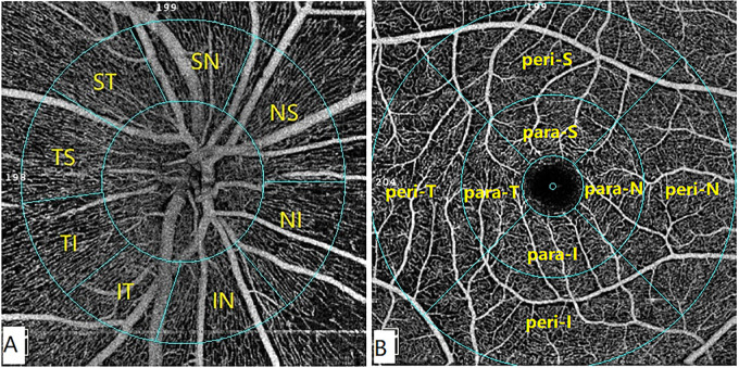
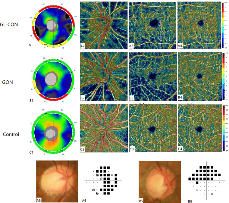
In this retrospective cross-sectional study, OCT scans were performed on 28 eyes of GL-CON, 34 eyes of GON, and 41control eyes to determine the pRNFL thickness, ganglion cell complex thickness, and cup/disc ratio. OCTA scans were conducted for 12 eyes of GL-CON, 15 eyes of GON, and 15 control eyes to measure the vessel density of the peripapillary and macular areas. Analysis of covariance was used to perform the comparisons, and the area under the curve was calculated.
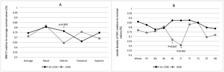
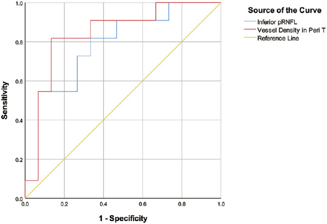
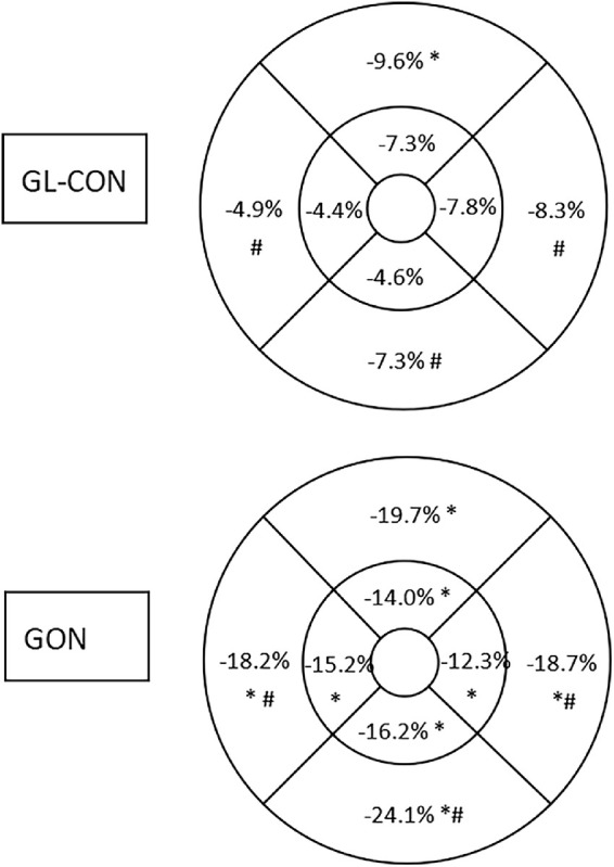
The GON eyes had a significantly thinner pRNFL in the inferior quadrant and greater vertical cup/disc ratio than the GL-CON eyes. In the radial peripapillary capillary segment, the vessel density of the GON in the inferior sectors was significantly lower than in the GL-CON. The superficial macular vessel density in the whole-image, peritemporal, perinasal, and peri-inferior sectors was significantly smaller in the GON group than in the GL-CON group. The best parameter for discriminating between GL-CON and GON was the superficial macular vessel density in the peritemporal sector.
GL-CON eyes showed a characteristic pattern of pRNFL and retinal microvascular changes.
GL-CON can be effectively distinguished from GON by detecting the alterations in the pRNFL and retinal microvasculature using OCT and OCTA.
通过比较光学相干断层扫描(OCT)和光学相干断层扫描血管造影(OCTA)的视盘周围视网膜神经纤维层(pRNFL)厚度和视网膜微血管,来区分伴青光眼样杯盘比(GL-CON)的压迫性视神经病变和青光眼性视神经病变(GON)。
在这项回顾性的横断面研究中,对 28 只 GL-CON 眼、34 只 GON 眼和 41 只对照眼进行 OCT 扫描,以确定 pRNFL 厚度、神经节细胞复合体厚度和杯盘比。对 12 只 GL-CON 眼、15 只 GON 眼和 15 只对照眼进行 OCTA 扫描,以测量视盘周围和黄斑区的血管密度。采用协方差分析进行比较,并计算曲线下面积。
GON 眼的下象限 pRNFL 明显变薄,垂直杯盘比大于 GL-CON 眼。在放射状视盘毛细血管节段,GON 的下象限血管密度明显低于 GL-CON。GON 组的全像、颞侧、鼻侧和下象限的浅层黄斑血管密度明显小于 GL-CON 组。区分 GL-CON 和 GON 的最佳参数是颞侧全像的浅层黄斑血管密度。
GL-CON 眼表现出 pRNFL 和视网膜微血管变化的特征模式。
翻译的准确性和流畅度取决于许多因素,包括源文本的质量、语法和词汇的复杂性、翻译的目的和受众等。因此,虽然我已经尽力提供准确和流畅的翻译,但仍有可能存在一些不准确或不自然的表达方式。如果你发现任何问题或有任何改进的建议,请随时告诉我。