Orford C R, Gardner D L, O'Connor P, Bates G, Swallow J J, Brito-Babapulle L A
Department of Histopathology, University Hospital of South Manchester.
J Anat. 1986 Oct;148:233-44.
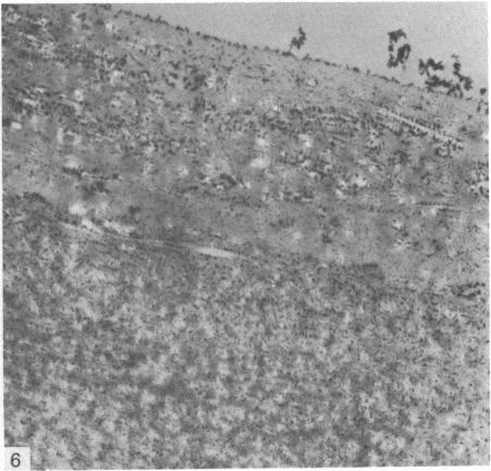
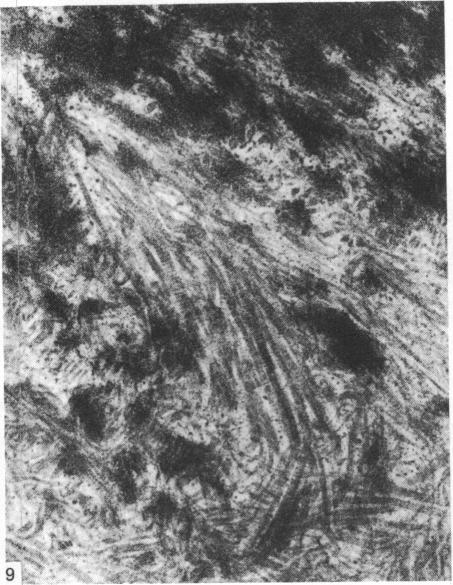
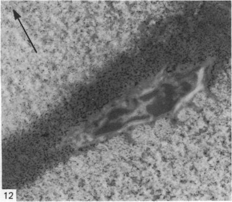
The cationic dye, cupromeronic blue, has been used in a critical electrolyte concentration technique to analyse the ultrastructural changes in cartilage matrix glycosaminoglycans which occur in the dog anterior cruciate ligament division model of osteoarthrosis. Amorphous material appearing at the articular surface of cartilage from the stifle joints of animals subjected to open surgical division of the anterior cruciate ligament has been shown not to comprise glycosaminoglycan. The nature of this material is unknown, but it appears to replace the surface lamina of normal cartilage. It may therefore affect the mechanical properties of the superficial cartilage. The pericellular matrix around single chondrocytes or separating pairs of chondrocytes becomes enriched with sulphated glycosaminoglycan as a response to ligament section. This material is thought to be newly synthesised and secreted and reflects the increased cellular activity resulting from surgically induced canine joint disease.
阳离子染料铜铬黑蓝已被用于一种临界电解质浓度技术,以分析骨关节炎犬前交叉韧带断裂模型中软骨基质糖胺聚糖的超微结构变化。在接受前交叉韧带开放性手术切断的动物膝关节软骨关节表面出现的无定形物质已被证明不包含糖胺聚糖。这种物质的性质尚不清楚,但它似乎取代了正常软骨的表面层。因此,它可能会影响表层软骨的力学性能。作为对韧带切断的反应,单个软骨细胞周围或成对软骨细胞之间的细胞周基质中硫酸化糖胺聚糖变得丰富。这种物质被认为是新合成并分泌的,反映了手术诱导的犬类关节疾病导致的细胞活性增加。