• 文献检索
  • 文档翻译
  • 深度研究
  • 学术资讯
  • Suppr Zotero 插件Zotero 插件
  • 邀请有礼
  • 套餐&价格
  • 历史记录
应用&插件
Suppr Zotero 插件Zotero 插件浏览器插件Mac 客户端Windows 客户端微信小程序
定价
高级版会员购买积分包购买API积分包
服务
文献检索文档翻译深度研究API 文档MCP 服务
关于我们
关于 Suppr公司介绍联系我们用户协议隐私条款
关注我们

Suppr 超能文献

核心技术专利:CN118964589B侵权必究
粤ICP备2023148730 号-1Suppr @ 2026

文献检索

告别复杂PubMed语法,用中文像聊天一样搜索,搜遍4000万医学文献。AI智能推荐,让科研检索更轻松。

立即免费搜索

文件翻译

保留排版,准确专业,支持PDF/Word/PPT等文件格式,支持 12+语言互译。

免费翻译文档

深度研究

AI帮你快速写综述,25分钟生成高质量综述,智能提取关键信息,辅助科研写作。

立即免费体验

颈外静脉的解剖变异:图像学与批判性综述。

Anatomical Variations of the External Jugular Vein: A Pictorial and Critical Review.

机构信息

Division of Anatomy, Faculty of Stomatology, "Carol Davila" University of Medicine and Pharmacy, 020021 Bucharest, Romania.

Division of Neurosurgery, Department 6-Clinical Neurosciences, Faculty of Medicine, "Carol Davila" University of Medicine and Pharmacy, 020021 Bucharest, Romania.

出版信息

Medicina (Kaunas). 2023 Mar 21;59(3):622. doi: 10.3390/medicina59030622.

DOI:10.3390/medicina59030622
PMID:36984623
原文链接:https://pmc.ncbi.nlm.nih.gov/articles/PMC10052824/
Abstract

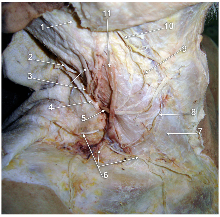
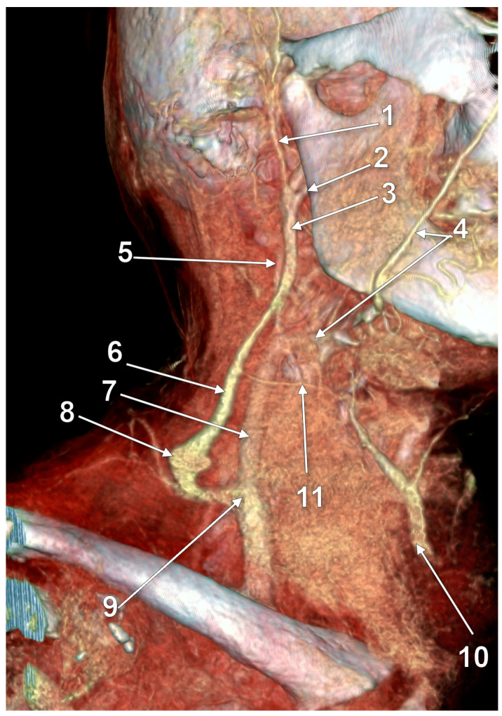
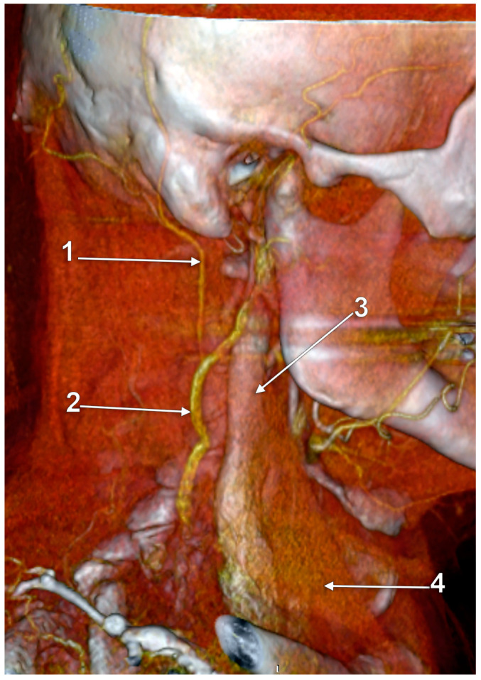
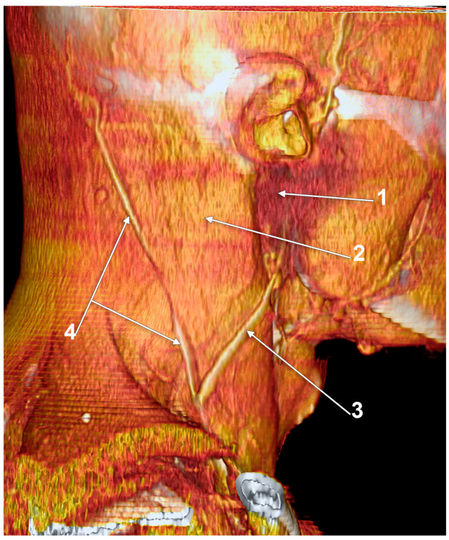
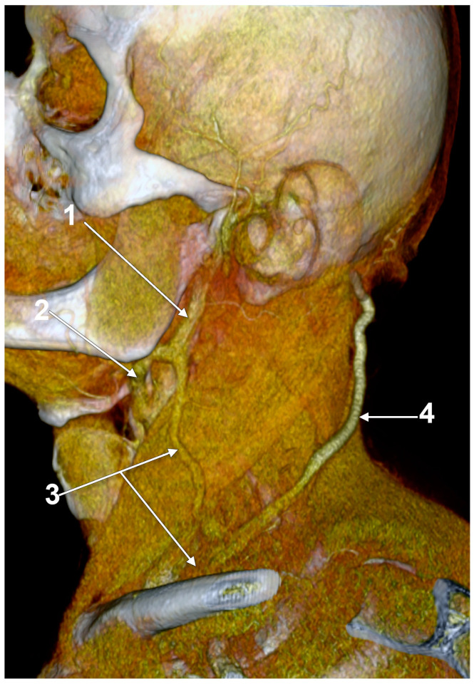
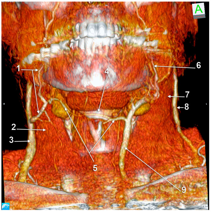
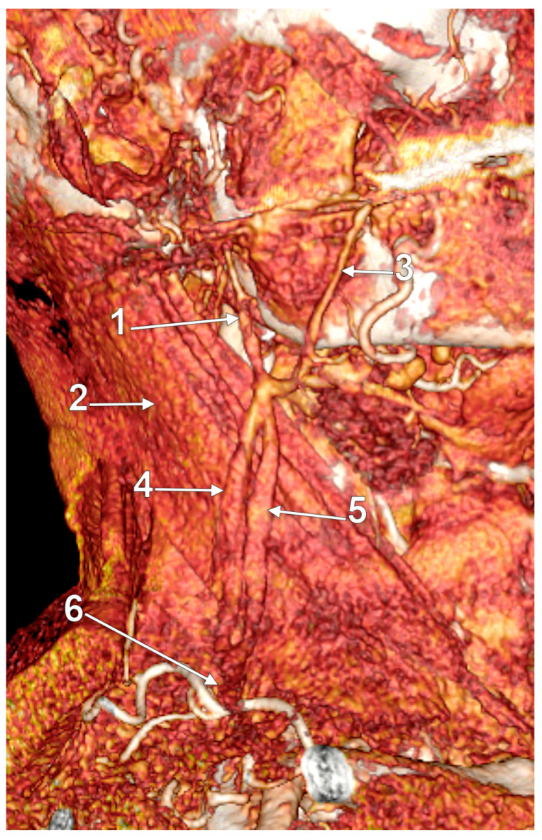
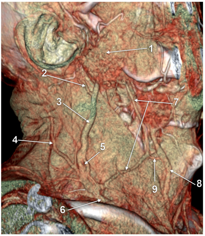
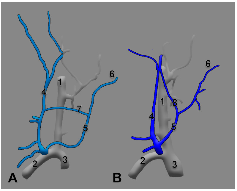
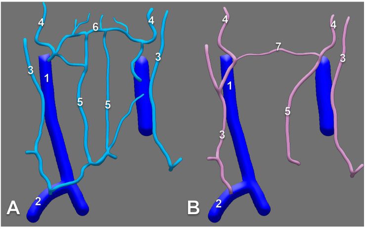
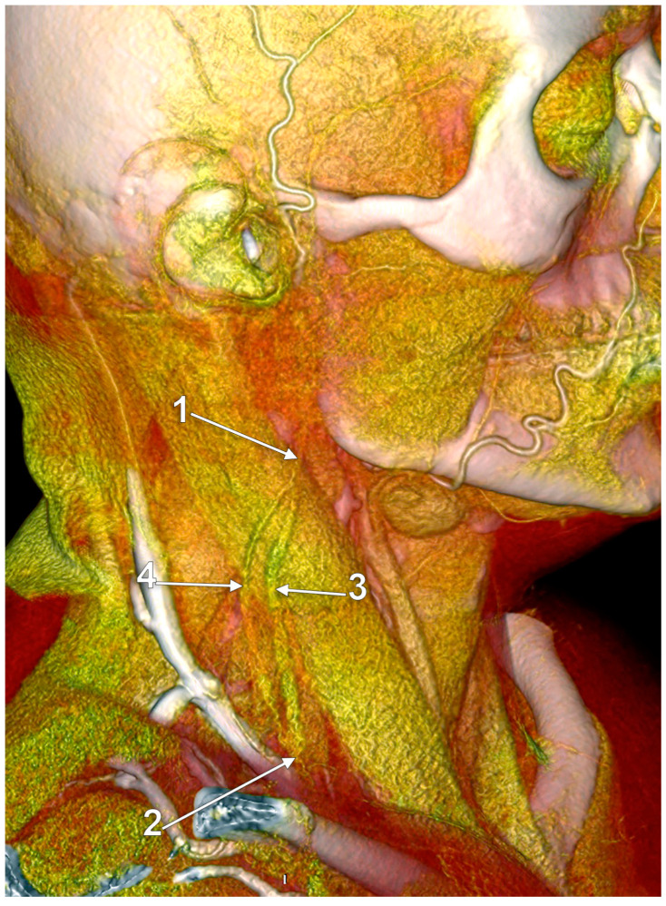
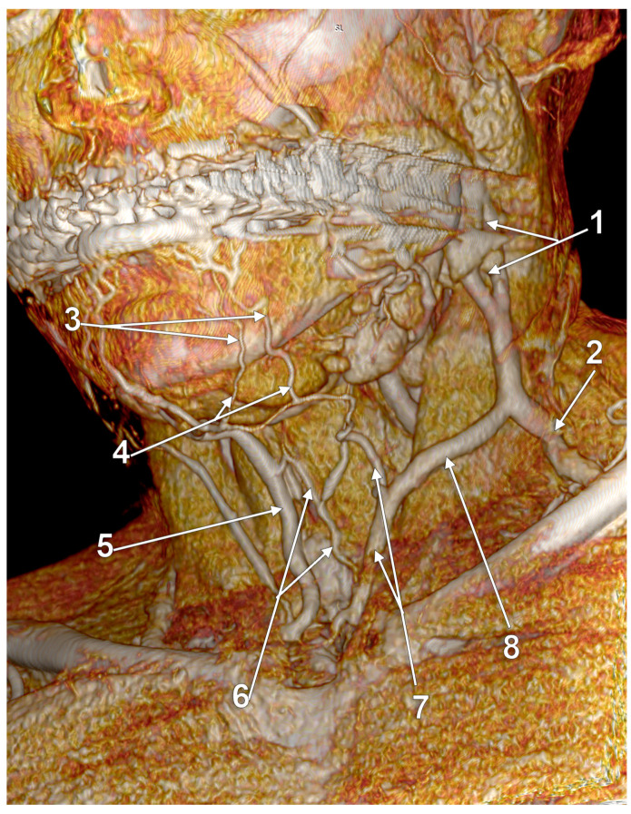
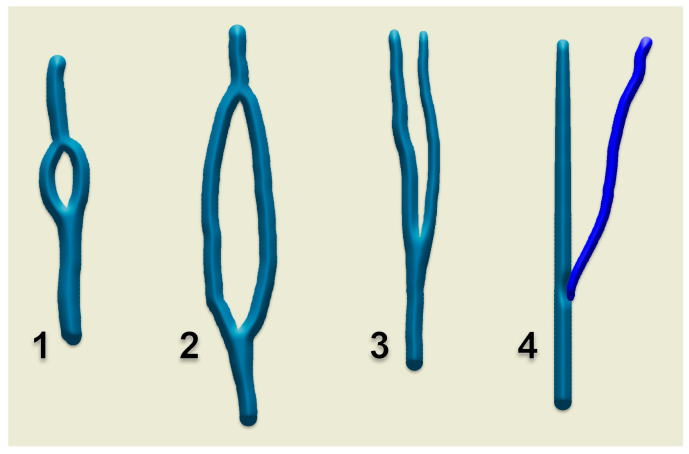
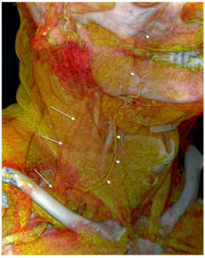
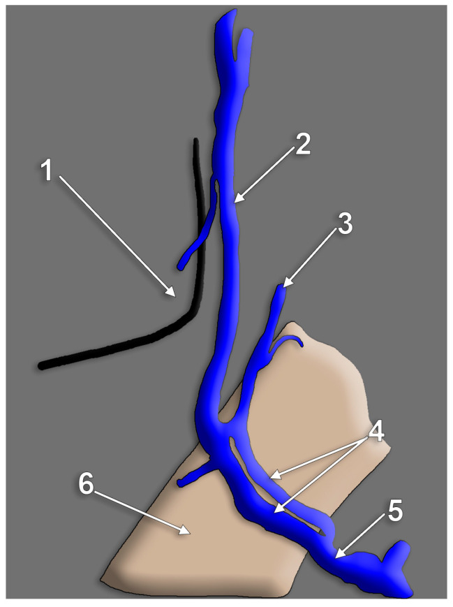
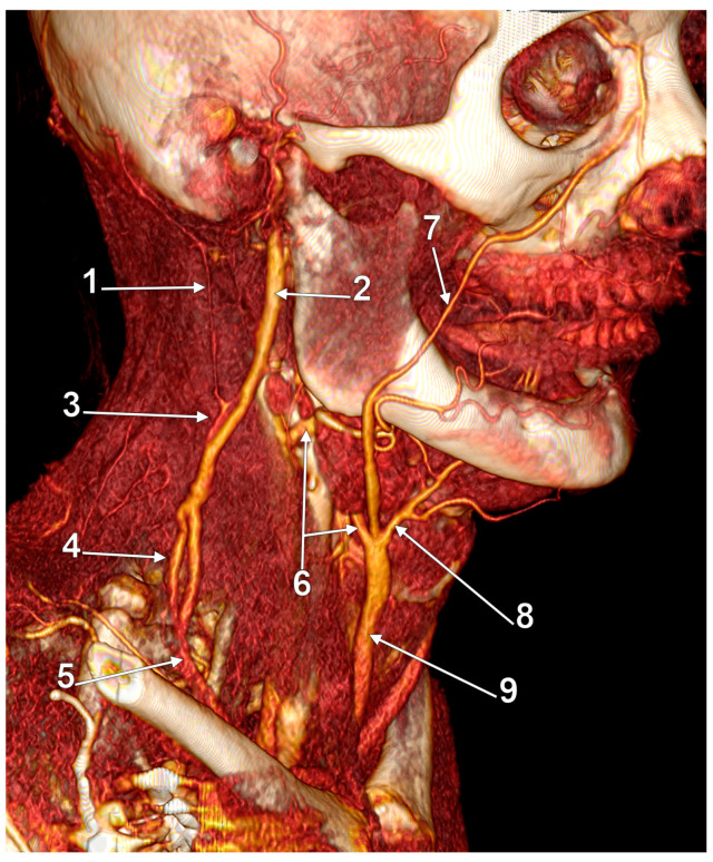
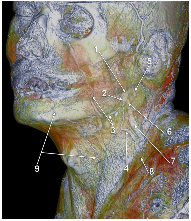
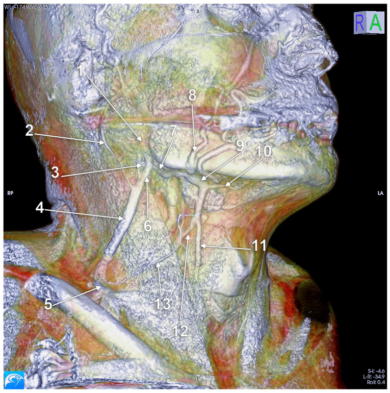
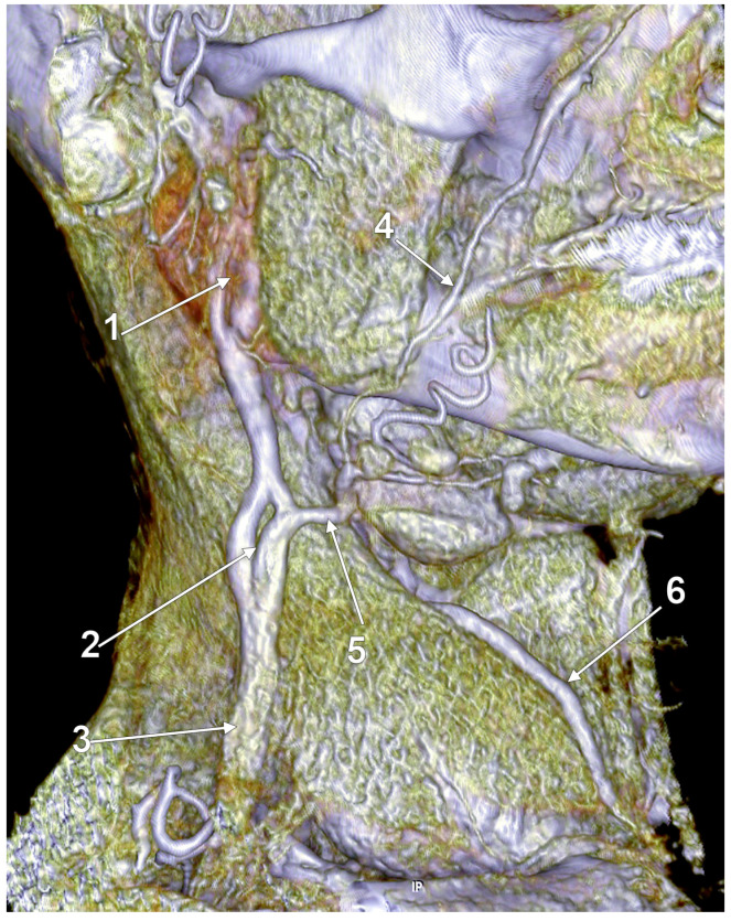
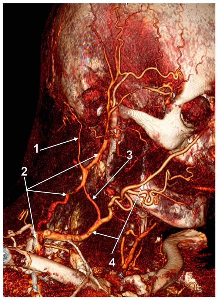
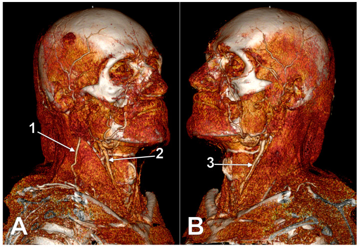
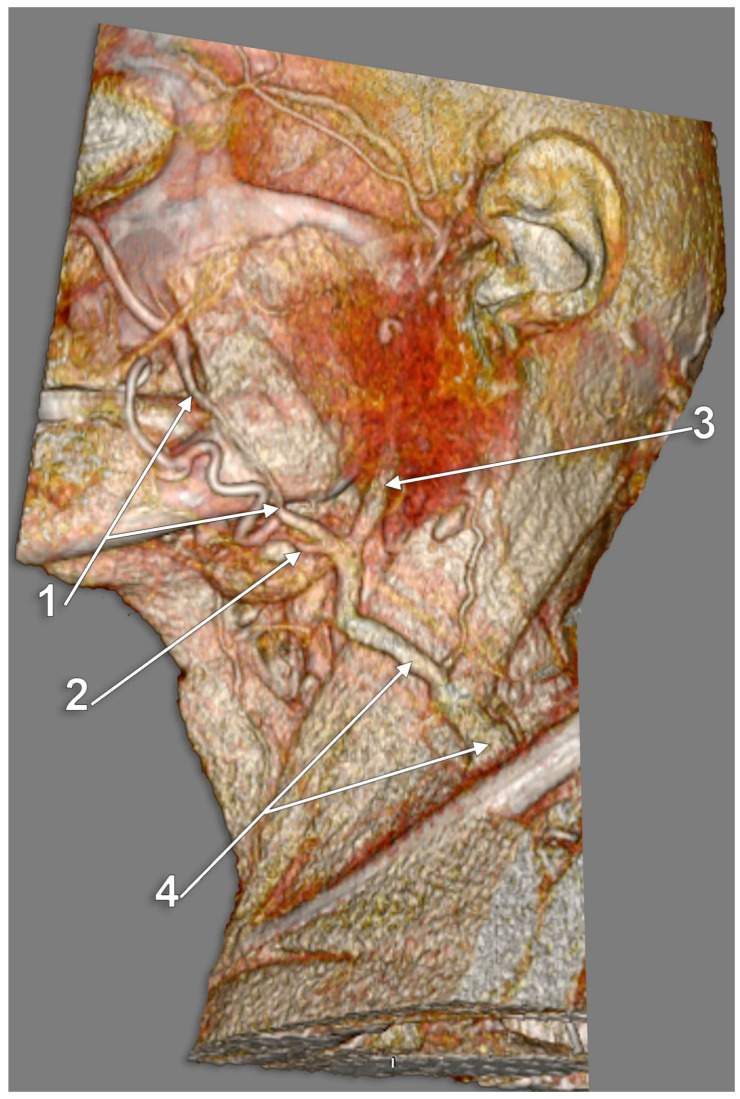
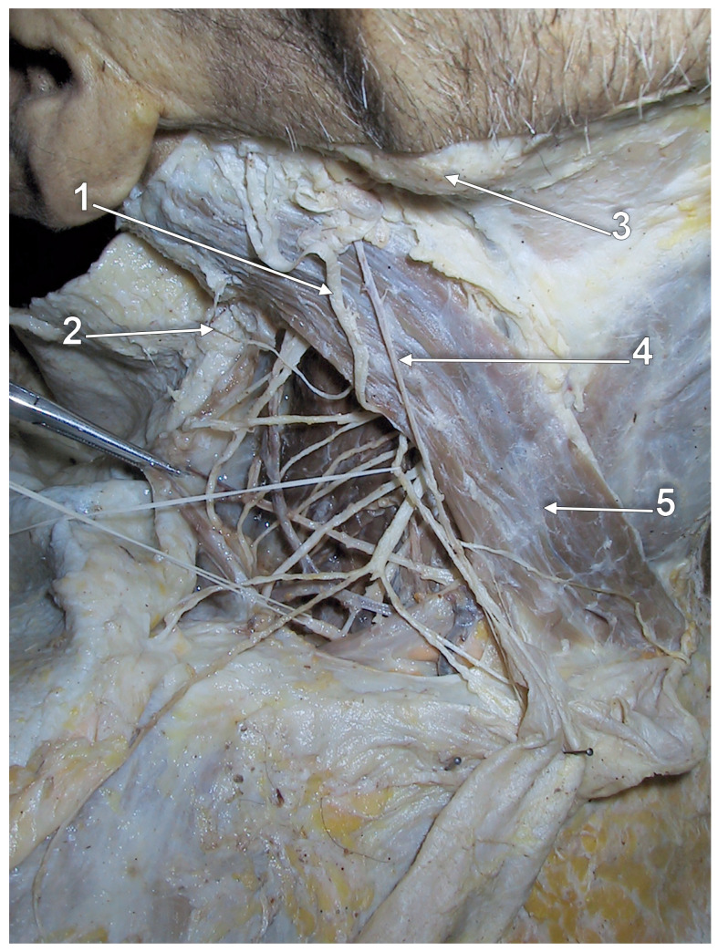
(1) : The external jugular vein (EJV) descends on the sternocleidomastoid muscle to drain deep into the subclavian vein. Anatomical variations of the EJV are relevant for identification of the greater auricular nerve, flap design and preparation, or EJV cannulation. (2) : Different publications were comprehensively reviewed. Dissections and three-dimensional volume renderings of peculiar cases were used to sample the review. (3) : Different anatomical possibilities of the EJV were critically reviewed and documented: fenestrations and double fenestrations, true or false duplications, triplication, absence, aberrant origin or course, or bifurcation. Tributaries of the EJV, such as the facial and posterior external jugular veins, are discussed. The internal jugular vein termination of the EJV is also presented. (4) : Care should be taken when different morphological features of the EJV are encountered or reported.

摘要

(1):颈外静脉(EJV)沿胸锁乳突肌下降,深入注入锁骨下静脉。EJV 的解剖变异与耳大神经的识别、皮瓣设计和准备或 EJV 插管有关。

(2):对不同的出版物进行了全面综述。对特殊病例的解剖和三维容积渲染进行了采样,以进行综述。

(3):对 EJV 的不同解剖可能性进行了批判性回顾和记录:窗孔和双窗孔、真性或假性重复、三重、缺失、异常起源或走行、或分叉。还讨论了 EJV 的属支,如面静脉和后颈外静脉。EJV 汇入颈内静脉的终点也有所呈现。

(4):当遇到或报告 EJV 的不同形态特征时,应加以注意。

https://cdn.ncbi.nlm.nih.gov/pmc/blobs/a89e/10052824/1c94fec7ca37/medicina-59-00622-g024.jpg
https://cdn.ncbi.nlm.nih.gov/pmc/blobs/a89e/10052824/f4830abcbaa4/medicina-59-00622-g001.jpg
https://cdn.ncbi.nlm.nih.gov/pmc/blobs/a89e/10052824/d06e688d029d/medicina-59-00622-g002.jpg
https://cdn.ncbi.nlm.nih.gov/pmc/blobs/a89e/10052824/be96ce571fdc/medicina-59-00622-g003.jpg
https://cdn.ncbi.nlm.nih.gov/pmc/blobs/a89e/10052824/109cccff6ca8/medicina-59-00622-g004.jpg
https://cdn.ncbi.nlm.nih.gov/pmc/blobs/a89e/10052824/f1c3477adf59/medicina-59-00622-g005.jpg
https://cdn.ncbi.nlm.nih.gov/pmc/blobs/a89e/10052824/af0f8261a47b/medicina-59-00622-g006.jpg
https://cdn.ncbi.nlm.nih.gov/pmc/blobs/a89e/10052824/81193e11d371/medicina-59-00622-g007.jpg
https://cdn.ncbi.nlm.nih.gov/pmc/blobs/a89e/10052824/c4c528e92d16/medicina-59-00622-g008.jpg
https://cdn.ncbi.nlm.nih.gov/pmc/blobs/a89e/10052824/ab69dd7f4dff/medicina-59-00622-g009.jpg
https://cdn.ncbi.nlm.nih.gov/pmc/blobs/a89e/10052824/5b7a92208b3a/medicina-59-00622-g010.jpg
https://cdn.ncbi.nlm.nih.gov/pmc/blobs/a89e/10052824/1dafa2726704/medicina-59-00622-g011.jpg
https://cdn.ncbi.nlm.nih.gov/pmc/blobs/a89e/10052824/5685362a8fa7/medicina-59-00622-g012.jpg
https://cdn.ncbi.nlm.nih.gov/pmc/blobs/a89e/10052824/76343039e8d1/medicina-59-00622-g013.jpg
https://cdn.ncbi.nlm.nih.gov/pmc/blobs/a89e/10052824/e8352f5df8d9/medicina-59-00622-g014.jpg
https://cdn.ncbi.nlm.nih.gov/pmc/blobs/a89e/10052824/1006a1a629d3/medicina-59-00622-g015.jpg
https://cdn.ncbi.nlm.nih.gov/pmc/blobs/a89e/10052824/64f542b9815b/medicina-59-00622-g016.jpg
https://cdn.ncbi.nlm.nih.gov/pmc/blobs/a89e/10052824/e9d06e1f8110/medicina-59-00622-g017.jpg
https://cdn.ncbi.nlm.nih.gov/pmc/blobs/a89e/10052824/3a180dd34597/medicina-59-00622-g018.jpg
https://cdn.ncbi.nlm.nih.gov/pmc/blobs/a89e/10052824/33a2541d8eb2/medicina-59-00622-g019.jpg
https://cdn.ncbi.nlm.nih.gov/pmc/blobs/a89e/10052824/d0e58ac36d49/medicina-59-00622-g020.jpg
https://cdn.ncbi.nlm.nih.gov/pmc/blobs/a89e/10052824/7f8b2b0b9431/medicina-59-00622-g021.jpg
https://cdn.ncbi.nlm.nih.gov/pmc/blobs/a89e/10052824/042f921af606/medicina-59-00622-g022.jpg
https://cdn.ncbi.nlm.nih.gov/pmc/blobs/a89e/10052824/3274fb59cb06/medicina-59-00622-g023.jpg
https://cdn.ncbi.nlm.nih.gov/pmc/blobs/a89e/10052824/1c94fec7ca37/medicina-59-00622-g024.jpg

相似文献

1
Anatomical Variations of the External Jugular Vein: A Pictorial and Critical Review.颈外静脉的解剖变异:图像学与批判性综述。
Medicina (Kaunas). 2023 Mar 21;59(3):622. doi: 10.3390/medicina59030622.
2
Duplication of the external jugular vein: a language barrier of database search in classic anatomical studies.颈外静脉重复:经典解剖学研究中数据库检索的语言障碍。
Surg Radiol Anat. 2021 Oct;43(10):1721-1728. doi: 10.1007/s00276-021-02717-6. Epub 2021 Feb 23.
3
Facial vein draining into external jugular vein in humans: its variations, phylogenetic retention and clinical relevance.人类面静脉汇入颈外静脉:其变异、系统发育保留及临床意义。
Surg Radiol Anat. 2003 Apr;25(1):36-41. doi: 10.1007/s00276-002-0080-z. Epub 2003 Jan 21.
4
Review of the Variations of the Superficial Veins of the Neck.颈部浅静脉变异的综述
Cureus. 2018 Jun 18;10(6):e2826. doi: 10.7759/cureus.2826.
5
Facial vein terminating in the external jugular vein. An embryologic interpretation.面静脉汇入颈外静脉。胚胎学解释。
Surg Radiol Anat. 1997;19(2):73-7.
6
An unilateral double fenestration of the right external jugular vein: a rare variant.右侧颈外静脉单侧双重窗孔:一种罕见的变异。
Surg Radiol Anat. 2024 Nov;46(11):1807-1810. doi: 10.1007/s00276-024-03473-z. Epub 2024 Aug 30.
7
Computed tomographic characteristics of anatomical variations of external and internal jugular veins in dogs.犬颈外静脉和颈内静脉解剖变异的 CT 特征。
Vet Radiol Ultrasound. 2024 Nov;65(6):727-734. doi: 10.1111/vru.13419. Epub 2024 Aug 19.
8
The importance of the angulation and termination of external jugular vein in central venous catheterization in newborn.新生儿中心静脉置管中外颈静脉角度和终点的重要性。
Okajimas Folia Anat Jpn. 1996 Aug;73(2-3):155-9. doi: 10.2535/ofaj1936.73.2-3_155.
9
Unilateral external jugular vein fenestration with variant anatomy of the retromandibular and facial vein.伴有下颌后静脉和面部静脉解剖变异的单侧颈外静脉开窗术
Anat Cell Biol. 2020 Mar;53(1):117-120. doi: 10.5115/acb.19.172. Epub 2020 Mar 3.
10
Central venous access via external jugular vein with CT-venography using a multidetector helical 16-section CT.通过颈外静脉进行中心静脉置管,并使用16层螺旋CT进行CT静脉造影。
J Invest Surg. 2014 Jun;27(3):176-82. doi: 10.3109/08941939.2013.865818. Epub 2013 Dec 19.

引用本文的文献

1
A Rare Case of Paired Congenital Cervical Aneurysms in a Communicating Vein: Clinical and Imaging Findings in a Pediatric Patient.一例罕见的交通静脉内双侧先天性颈动脉瘤病例:一名儿科患者的临床及影像学表现
Pathophysiology. 2025 Jun 6;32(2):25. doi: 10.3390/pathophysiology32020025.
2
Tunneled Dialysis Catheter Insertion in External Jugular Vein by Nephrologists.肾脏病学家在外颈静脉进行带隧道透析导管置入术。
Indian J Nephrol. 2025 Jan-Feb;35(1):46-52. doi: 10.25259/ijn_511_23. Epub 2024 Jul 8.
3
Anatomical variations of the external jugular veins and collaterals incidentally diagnosed with computed tomography in Shih Tzu dogs.

本文引用的文献

1
The transcerebral laterocavernous vein, a form of persisting primitive tentorial sinus.大脑外侧岩下窦,一种持续存在的原始天幕窦形式。
Surg Radiol Anat. 2022 Nov;44(11):1471-1474. doi: 10.1007/s00276-022-03038-y. Epub 2022 Oct 27.
2
Transcallosal and Pericallosal Courses of the Anterior Cerebral Artery.大脑前动脉的胼周和胼缘动脉。
Medicina (Kaunas). 2022 Sep 28;58(10):1365. doi: 10.3390/medicina58101365.
3
A New Classification of the Anatomical Variations of Labbé's Inferior Anastomotic Vein.拉贝下吻合静脉解剖变异的新分类。
西施犬经计算机断层扫描偶然诊断出的颈外静脉和侧支的解剖变异。
Front Vet Sci. 2024 Oct 17;11:1464750. doi: 10.3389/fvets.2024.1464750. eCollection 2024.
4
Isolated external jugular thrombophlebitis secondary to acute pharyngitis: a case report and a review of the literature.孤立性颈外静脉血栓性静脉炎继发于急性咽炎:病例报告及文献复习。
Ital J Pediatr. 2024 Sep 16;50(1):179. doi: 10.1186/s13052-024-01760-4.
5
An Ectopic External Jugular Vein Draining into the Axillary Vein: a Rare Anatomical Variation with Clinical Implications.一条异位的颈外静脉引流至腋静脉:一种具有临床意义的罕见解剖变异。
Acta Med Acad. 2024 Dec;53(3):309-313. doi: 10.5644/ama2006-124.441.
6
Cannulation via the external jugular vein--An alternative to conventional peripherally inserted central catheterisation for paediatric patients.经颈外静脉置管术——儿科患者传统外周中心静脉置管的替代方法。
BMC Pediatr. 2023 Nov 18;23(1):579. doi: 10.1186/s12887-023-04403-5.
Tomography. 2022 Aug 30;8(5):2182-2192. doi: 10.3390/tomography8050183.
4
Evaluation of the External Jugular Vein Overlying the Sternocleidomastoid Muscle as Venous Lymph-Node Flap.评估胸锁乳突肌表面的颈外静脉作为静脉淋巴结瓣的情况。
J Clin Med. 2022 Mar 25;11(7):1812. doi: 10.3390/jcm11071812.
5
Launay's External Carotid Vein.朗厄氏颈外静脉。
Medicina (Kaunas). 2021 Sep 18;57(9):985. doi: 10.3390/medicina57090985.
6
Thrombosed external jugular vein aneurysm mimics to a branchial cyst: A novel case from Iraq and review of the literature.血栓形成的颈外静脉动脉瘤酷似鳃裂囊肿:来自伊拉克的一例新病例及文献综述。
Ann Med Surg (Lond). 2021 Jun 29;67:102533. doi: 10.1016/j.amsu.2021.102533. eCollection 2021 Jul.
7
Duplicated transverse cervical nerve and external jugular vein.副横颈神经和颈外静脉。
Anat Cell Biol. 2021 Sep 30;54(3):404-406. doi: 10.5115/acb.21.071.
8
Duplication of the external jugular vein: a language barrier of database search in classic anatomical studies.颈外静脉重复:经典解剖学研究中数据库检索的语言障碍。
Surg Radiol Anat. 2021 Oct;43(10):1721-1728. doi: 10.1007/s00276-021-02717-6. Epub 2021 Feb 23.
9
Supraclavicular nerve entrapment by the external jugular vein: An unreported finding.锁骨上神经被颈外静脉卡压:一种未报道的发现。
Morphologie. 2022 Feb;106(352):52-55. doi: 10.1016/j.morpho.2020.12.006. Epub 2021 Jan 19.
10
External jugular vein pierced by supraclavicular branches in a neonatal cadaver: a case report.新生儿尸体中锁骨上支穿刺颈外静脉:1 例报告。
Anat Sci Int. 2021 Sep;96(4):564-567. doi: 10.1007/s12565-020-00596-2. Epub 2021 Jan 8.