Eunice Kennedy Shriver National Institute of Child Health and Human Development (NICHD), National Institutes of Health, Bethesda, MD, USA.
Department of Human Development and Quantitative Methodology, University of Maryland, College Park, MD, USA.
Sci Rep. 2023 Mar 29;13(1):5151. doi: 10.1038/s41598-023-31609-5.
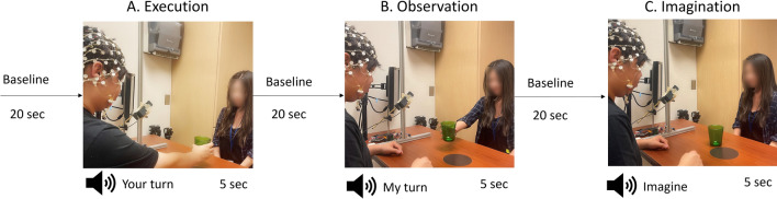
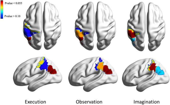
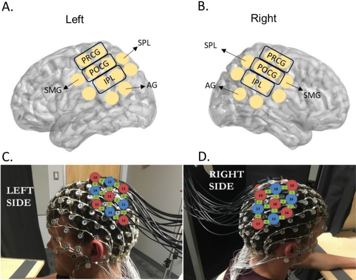
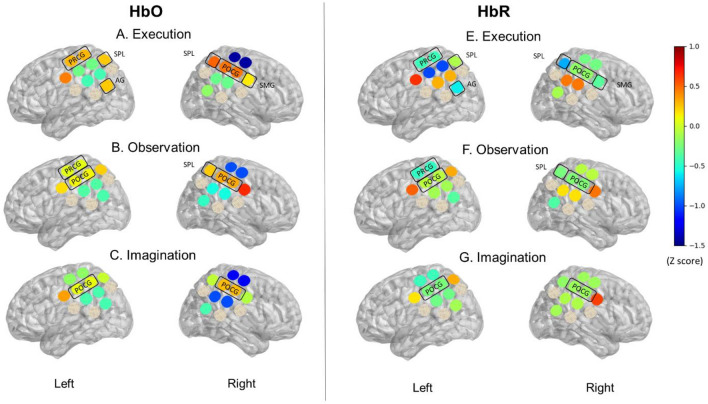
Motor execution, observation, and imagery are important skills used in motor learning and rehabilitation. The neural mechanisms underlying these cognitive-motor processes are still poorly understood. We used a simultaneous recording of functional near-infrared spectroscopy (fNIRS) and electroencephalogram (EEG) to elucidate the differences in neural activity across three conditions requiring these processes. Additionally, we used a new method called structured sparse multiset Canonical Correlation Analysis (ssmCCA) to fuse the fNIRS and EEG data and determine the brain regions of neural activity consistently detected by both modalities. Unimodal analyses revealed differentiated activation between conditions; however, the activated regions did not fully overlap across the two modalities (fNIRS: left angular gyrus, right supramarginal gyrus, as well as right superior and inferior parietal lobes; EEG: bilateral central, right frontal, and parietal). These discrepancies might be because fNIRS and EEG detect different signals. Using fused fNIRS-EEG data, we consistently found activation over the left inferior parietal lobe, superior marginal gyrus, and post-central gyrus during all three conditions, suggesting that our multimodal approach identifies a shared neural region associated with the Action Observation Network (AON). This study highlights the strengths of using the multimodal fNIRS-EEG fusion technique for studying AON. Neural researchers should consider using the multimodal approach to validate their findings.
运动执行、观察和意象是运动学习和康复中使用的重要技能。这些认知-运动过程的神经机制仍知之甚少。我们使用功能近红外光谱 (fNIRS) 和脑电图 (EEG) 的同步记录来阐明这三个需要这些过程的条件下的神经活动差异。此外,我们使用一种称为结构稀疏多集典型相关分析 (ssmCCA) 的新方法来融合 fNIRS 和 EEG 数据,并确定两种模态一致检测到的神经活动的大脑区域。单模态分析显示条件之间存在差异激活;然而,激活区域在两种模态之间并未完全重叠(fNIRS:左角回、右缘上回以及右顶叶和下顶叶;EEG:双侧中央、右额和顶叶)。这些差异可能是因为 fNIRS 和 EEG 检测到不同的信号。使用融合的 fNIRS-EEG 数据,我们在所有三种条件下都一致地发现左顶下叶、上缘回和后中央回的激活,这表明我们的多模态方法确定了与动作观察网络 (AON) 相关的共享神经区域。这项研究强调了使用多模态 fNIRS-EEG 融合技术研究 AON 的优势。神经研究人员应该考虑使用多模态方法来验证他们的发现。