• 文献检索
  • 文档翻译
  • 深度研究
  • 学术资讯
  • Suppr Zotero 插件Zotero 插件
  • 邀请有礼
  • 套餐&价格
  • 历史记录
应用&插件
Suppr Zotero 插件Zotero 插件浏览器插件Mac 客户端Windows 客户端微信小程序
定价
高级版会员购买积分包购买API积分包
服务
文献检索文档翻译深度研究API 文档MCP 服务
关于我们
关于 Suppr公司介绍联系我们用户协议隐私条款
关注我们

Suppr 超能文献

核心技术专利:CN118964589B侵权必究
粤ICP备2023148730 号-1Suppr @ 2026

文献检索

告别复杂PubMed语法,用中文像聊天一样搜索,搜遍4000万医学文献。AI智能推荐,让科研检索更轻松。

立即免费搜索

文件翻译

保留排版,准确专业,支持PDF/Word/PPT等文件格式,支持 12+语言互译。

免费翻译文档

深度研究

AI帮你快速写综述,25分钟生成高质量综述,智能提取关键信息,辅助科研写作。

立即免费体验

单细胞 RNA 测序显示心脏细胞间通讯在糖尿病心肌病心肌纤维化中的驱动因素。

Single-cell RNA-seq of heart reveals intercellular communication drivers of myocardial fibrosis in diabetic cardiomyopathy.

机构信息

Cyrus Tang Hematology Center, Soochow University, Suzhou, China.

Institute of Clinical Medicine Research, Suzhou Science & Technology Town Hospital, Gusu School, Nanjing Medical University, Suzhou, China.

出版信息

Elife. 2023 Apr 3;12:e80479. doi: 10.7554/eLife.80479.

DOI:10.7554/eLife.80479
PMID:37010266
原文链接:https://pmc.ncbi.nlm.nih.gov/articles/PMC10110238/
Abstract

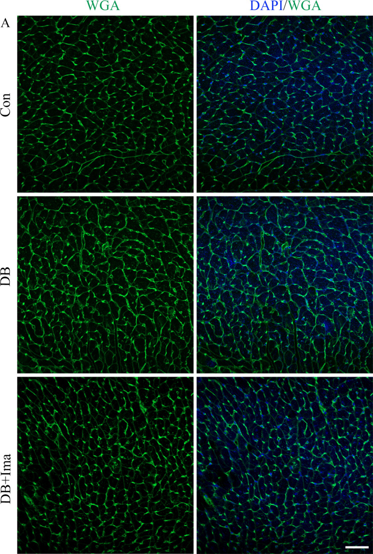
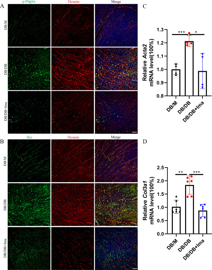
Myocardial fibrosis is the characteristic pathology of diabetes-induced cardiomyopathy. Therefore, an in-depth study of cardiac heterogeneity and cell-to-cell interactions can help elucidate the pathogenesis of diabetic myocardial fibrosis and identify treatment targets for the treatment of this disease. In this study, we investigated intercellular communication drivers of myocardial fibrosis in mouse heart with high-fat-diet/streptozotocin-induced diabetes at single-cell resolution. Intercellular and protein-protein interaction networks of fibroblasts and macrophages, endothelial cells, as well as fibroblasts and epicardial cells revealed critical changes in ligand-receptor interactions such as Pdgf(s)-Pdgfra and Efemp1-Egfr, which promote the development of a profibrotic microenvironment during the progression of and confirmed that the specific inhibition of the Pdgfra axis could significantly improve diabetic myocardial fibrosis. We also identified phenotypically distinct and fibroblast subpopulations associated with pathological extracellular matrix remodeling, of which the fibroblasts were found to be the most profibrogenic under diabetic conditions. Finally, we validated the role of the hub gene-mediated intercellular communication drivers of diabetic myocardial fibrosis in fibroblasts, and confirmed the results through AAV9-mediated knockdown in the heart of diabetic mice. In summary, cardiac cell mapping provides novel insights into intercellular communication drivers involved in pathological extracellular matrix remodeling during diabetic myocardial fibrosis.

摘要

心肌纤维化是糖尿病性心肌病的特征性病理学改变。因此,深入研究心脏异质性和细胞间相互作用有助于阐明糖尿病性心肌纤维化的发病机制,并确定治疗这种疾病的治疗靶点。在这项研究中,我们在单细胞分辨率下研究了高脂肪饮食/链脲佐菌素诱导的糖尿病小鼠心脏中细胞间通讯驱动心肌纤维化的机制。成纤维细胞和巨噬细胞、内皮细胞以及成纤维细胞和心外膜细胞的细胞间和蛋白-蛋白相互作用网络揭示了配体-受体相互作用的关键变化,如 Pdgf(s)-Pdgfra 和 Efemp1-Egfr,这些变化促进了糖尿病心肌纤维化进展过程中促纤维化微环境的发展,并证实了 Pdgfra 轴的特异性抑制可显著改善糖尿病性心肌纤维化。我们还鉴定了与病理性细胞外基质重塑相关的表型不同的成纤维细胞亚群,其中在糖尿病条件下, 成纤维细胞被发现是最具成纤维原性的。最后,我们验证了糖尿病心肌纤维化中 成纤维细胞中 基因介导的细胞间通讯驱动因子的作用,并通过在糖尿病小鼠心脏中使用 AAV9 介导的 基因敲低来证实了这一结果。总之,心脏细胞图谱为糖尿病性心肌纤维化过程中涉及病理性细胞外基质重塑的细胞间通讯驱动因素提供了新的见解。

https://cdn.ncbi.nlm.nih.gov/pmc/blobs/0244/10110238/5da9301a81ec/elife-80479-sa2-fig1.jpg
https://cdn.ncbi.nlm.nih.gov/pmc/blobs/0244/10110238/b4050d93c18a/elife-80479-fig1.jpg
https://cdn.ncbi.nlm.nih.gov/pmc/blobs/0244/10110238/869f0e55e267/elife-80479-fig1-figsupp1.jpg
https://cdn.ncbi.nlm.nih.gov/pmc/blobs/0244/10110238/7f74a43e3c02/elife-80479-fig1-figsupp2.jpg
https://cdn.ncbi.nlm.nih.gov/pmc/blobs/0244/10110238/3af53eca800d/elife-80479-fig1-figsupp3.jpg
https://cdn.ncbi.nlm.nih.gov/pmc/blobs/0244/10110238/aa4183fc37a2/elife-80479-fig2.jpg
https://cdn.ncbi.nlm.nih.gov/pmc/blobs/0244/10110238/d866a854f71f/elife-80479-fig2-figsupp1.jpg
https://cdn.ncbi.nlm.nih.gov/pmc/blobs/0244/10110238/1d9c0ad4b18f/elife-80479-fig3.jpg
https://cdn.ncbi.nlm.nih.gov/pmc/blobs/0244/10110238/efd939f4e46f/elife-80479-fig3-figsupp1.jpg
https://cdn.ncbi.nlm.nih.gov/pmc/blobs/0244/10110238/b6eddfa30cee/elife-80479-fig3-figsupp2.jpg
https://cdn.ncbi.nlm.nih.gov/pmc/blobs/0244/10110238/3d12cc50d7d8/elife-80479-fig3-figsupp3.jpg
https://cdn.ncbi.nlm.nih.gov/pmc/blobs/0244/10110238/442ed25e45ff/elife-80479-fig3-figsupp4.jpg
https://cdn.ncbi.nlm.nih.gov/pmc/blobs/0244/10110238/993d68250d16/elife-80479-fig3-figsupp5.jpg
https://cdn.ncbi.nlm.nih.gov/pmc/blobs/0244/10110238/3d8012297680/elife-80479-fig4.jpg
https://cdn.ncbi.nlm.nih.gov/pmc/blobs/0244/10110238/01be40c7c5eb/elife-80479-fig5.jpg
https://cdn.ncbi.nlm.nih.gov/pmc/blobs/0244/10110238/186635ab7d59/elife-80479-fig6.jpg
https://cdn.ncbi.nlm.nih.gov/pmc/blobs/0244/10110238/a5c07541b98a/elife-80479-fig6-figsupp1.jpg
https://cdn.ncbi.nlm.nih.gov/pmc/blobs/0244/10110238/75a8e3b3db47/elife-80479-fig6-figsupp2.jpg
https://cdn.ncbi.nlm.nih.gov/pmc/blobs/0244/10110238/e7b80f987dcf/elife-80479-fig7.jpg
https://cdn.ncbi.nlm.nih.gov/pmc/blobs/0244/10110238/c122b442ef2b/elife-80479-fig7-figsupp1.jpg
https://cdn.ncbi.nlm.nih.gov/pmc/blobs/0244/10110238/c81eedaa14c9/elife-80479-fig8.jpg
https://cdn.ncbi.nlm.nih.gov/pmc/blobs/0244/10110238/a2553f77c137/elife-80479-fig8-figsupp1.jpg
https://cdn.ncbi.nlm.nih.gov/pmc/blobs/0244/10110238/5da9301a81ec/elife-80479-sa2-fig1.jpg

相似文献

1
Single-cell RNA-seq of heart reveals intercellular communication drivers of myocardial fibrosis in diabetic cardiomyopathy.单细胞 RNA 测序显示心脏细胞间通讯在糖尿病心肌病心肌纤维化中的驱动因素。
Elife. 2023 Apr 3;12:e80479. doi: 10.7554/eLife.80479.
2
Single-cell insights: pioneering an integrated atlas of chromatin accessibility and transcriptomic landscapes in diabetic cardiomyopathy.单细胞洞察:开创糖尿病性心肌病染色质可及性和转录组景观综合图谱。
Cardiovasc Diabetol. 2024 Apr 25;23(1):139. doi: 10.1186/s12933-024-02233-y.
3
High-Resolution Transcriptomic Profiling of the Heart During Chronic Stress Reveals Cellular Drivers of Cardiac Fibrosis and Hypertrophy.慢性应激过程中心脏的高分辨率转录组特征分析揭示了心脏纤维化和肥大的细胞驱动因素。
Circulation. 2020 Oct 13;142(15):1448-1463. doi: 10.1161/CIRCULATIONAHA.119.045115. Epub 2020 Jul 30.
4
DNA methyltransferase-1 inactivation of androgen receptor axis triggers homocysteine induced cardiac fibroblast autophagy in diabetic cardiac fibrosis.DNA 甲基转移酶 1 失活雄激素受体轴触发同型半胱氨酸诱导的糖尿病心脏纤维化中心脏成纤维细胞自噬。
Arch Biochem Biophys. 2020 Oct 15;692:108521. doi: 10.1016/j.abb.2020.108521. Epub 2020 Aug 12.
5
MiR-21-3p triggers cardiac fibroblasts pyroptosis in diabetic cardiac fibrosis via inhibiting androgen receptor.miR-21-3p 通过抑制雄激素受体触发糖尿病心脏纤维化中的心肌成纤维细胞焦亡。
Exp Cell Res. 2021 Feb 15;399(2):112464. doi: 10.1016/j.yexcr.2020.112464. Epub 2020 Dec 29.
6
Targeted inhibition of calpain reduces myocardial hypertrophy and fibrosis in mouse models of type 1 diabetes.靶向抑制钙蛋白酶可减少 1 型糖尿病小鼠模型的心肌肥大和纤维化。
Diabetes. 2011 Nov;60(11):2985-94. doi: 10.2337/db10-1333. Epub 2011 Sep 12.
7
Irisin inhibits high glucose-induced endothelial-to-mesenchymal transition and exerts a dose-dependent bidirectional effect on diabetic cardiomyopathy.鸢尾素抑制高糖诱导的内皮细胞向间充质细胞转化,并对糖尿病心肌病发挥剂量依赖性双向作用。
J Cell Mol Med. 2018 Feb;22(2):808-822. doi: 10.1111/jcmm.13360. Epub 2017 Oct 23.
8
Antifibrotic cardioprotection of berberine via downregulating myocardial IGF-1 receptor-regulated MMP-2/MMP-9 expression in diabetic rats.小檗碱通过下调糖尿病大鼠心肌 IGF-1 受体调节的 MMP-2/MMP-9 表达发挥抗纤维化心脏保护作用。
Am J Physiol Heart Circ Physiol. 2018 Oct 1;315(4):H802-H813. doi: 10.1152/ajpheart.00093.2018. Epub 2018 Jun 29.
9
EGFR inhibition protects cardiac damage and remodeling through attenuating oxidative stress in STZ-induced diabetic mouse model.表皮生长因子受体抑制通过减轻 STZ 诱导的糖尿病小鼠模型中的氧化应激来保护心脏损伤和重构。
J Mol Cell Cardiol. 2015 May;82:63-74. doi: 10.1016/j.yjmcc.2015.02.029. Epub 2015 Mar 7.
10
Inhibition of high-mobility group box 1 improves myocardial fibrosis and dysfunction in diabetic cardiomyopathy.高迁移率族蛋白 1 抑制可改善糖尿病心肌病中的心肌纤维化和功能障碍。
Int J Cardiol. 2014 Mar 1;172(1):202-12. doi: 10.1016/j.ijcard.2014.01.011. Epub 2014 Jan 21.

引用本文的文献

1
TET3 is a regulator and can be targeted for the intervention of myocardial fibrosis.TET3是一种调节因子,可作为心肌纤维化干预的靶点。
EMBO Mol Med. 2025 Sep 8. doi: 10.1038/s44321-025-00305-4.
2
Omics-based Approach Towards Macrophages: New Perspectives of Biology and Function in the Normal and Diseased Heart.基于组学的巨噬细胞研究方法:正常和患病心脏中生物学与功能的新视角
Int J Biol Sci. 2025 May 27;21(8):3666-3688. doi: 10.7150/ijbs.112061. eCollection 2025.
3
Dynamic Expression and Functional Implications of the Cell Polarity Gene, Dchs1, During Cardiac Development.

本文引用的文献

1
Single-cell transcriptomics reveals cell-type-specific diversification in human heart failure.单细胞转录组学揭示了人类心力衰竭中细胞类型特异性的多样化。
Nat Cardiovasc Res. 2022 Mar;1(3):263-280. doi: 10.1038/s44161-022-00028-6. Epub 2022 Mar 16.
2
Regulation of Transplanted Cell Homing by FGF1 and PDGFB after Doxorubicin Myocardial Injury.阿霉素心肌损伤后 FGF1 和 PDGFB 对移植细胞归巢的调节。
Cells. 2021 Nov 3;10(11):2998. doi: 10.3390/cells10112998.
3
Single-cell RNA-seq reveals cellular heterogeneity of mouse carotid artery under disturbed flow.
细胞极性基因Dchs1在心脏发育过程中的动态表达及功能意义
Cells. 2025 May 24;14(11):774. doi: 10.3390/cells14110774.
4
Integrative Single-Cell and Spatial Transcriptomics Reveal Functional and Spatial Heterogeneity of Atrial and Ventricular Cardiomyocytes in the Heart.整合单细胞和空间转录组学揭示心脏中心房和心室心肌细胞的功能及空间异质性
Mol Biotechnol. 2025 Jun 9. doi: 10.1007/s12033-025-01443-3.
5
Identification of metabolic pathways and serum biomarkers in diabetic cardiomyopathy using untargeted metabolomics.使用非靶向代谢组学鉴定糖尿病性心肌病中的代谢途径和血清生物标志物
Sci Rep. 2025 May 28;15(1):18718. doi: 10.1038/s41598-025-98753-y.
6
Inhibiting atrial natriuretic peptide clearance reduces myocardial fibrosis and improves cardiac function in diabetic rats.抑制心房利钠肽清除可减轻糖尿病大鼠的心肌纤维化并改善心脏功能。
Eur Heart J Open. 2025 Mar 19;5(2):oeaf031. doi: 10.1093/ehjopen/oeaf031. eCollection 2025 Mar.
7
PD-1 inhibition disrupts collagen homeostasis and aggravates cardiac dysfunction through endothelial-fibroblast crosstalk and EndMT.程序性死亡受体1(PD-1)抑制通过内皮细胞-成纤维细胞相互作用和内皮-间质转化破坏胶原蛋白稳态并加重心脏功能障碍。
Front Pharmacol. 2025 Mar 17;16:1549487. doi: 10.3389/fphar.2025.1549487. eCollection 2025.
8
Cardiac Fibrosis in the Multi-Omics Era: Implications for Heart Failure.多组学时代的心脏纤维化:对心力衰竭的影响
Circ Res. 2025 Mar 28;136(7):773-802. doi: 10.1161/CIRCRESAHA.124.325402. Epub 2025 Mar 27.
9
Application and Progression of Single-Cell RNA Sequencing in Diabetes Mellitus and Diabetes Complications.单细胞RNA测序在糖尿病及糖尿病并发症中的应用与进展
J Diabetes Res. 2025 Mar 18;2025:3248350. doi: 10.1155/jdr/3248350. eCollection 2025.
10
Cellular crosstalk in organotypic vasculature: mechanisms of diabetic cardiorenal complications and SGLT2i responses.器官型脉管系统中的细胞间相互作用:糖尿病心肾并发症的机制及钠-葡萄糖协同转运蛋白2抑制剂的反应
Cardiovasc Diabetol. 2025 Feb 26;24(1):90. doi: 10.1186/s12933-025-02655-2.
单细胞RNA测序揭示了小鼠颈动脉在紊乱血流状态下的细胞异质性。
Cell Death Discov. 2021 Jul 16;7(1):180. doi: 10.1038/s41420-021-00567-0.
4
Cardiac fibrosis.心肌纤维化。
Cardiovasc Res. 2021 May 25;117(6):1450-1488. doi: 10.1093/cvr/cvaa324.
5
Cells of the adult human heart.成人心脏细胞。
Nature. 2020 Dec;588(7838):466-472. doi: 10.1038/s41586-020-2797-4. Epub 2020 Sep 24.
6
High-Resolution Transcriptomic Profiling of the Heart During Chronic Stress Reveals Cellular Drivers of Cardiac Fibrosis and Hypertrophy.慢性应激过程中心脏的高分辨率转录组特征分析揭示了心脏纤维化和肥大的细胞驱动因素。
Circulation. 2020 Oct 13;142(15):1448-1463. doi: 10.1161/CIRCULATIONAHA.119.045115. Epub 2020 Jul 30.
7
RAGE impairs murine diabetic atherosclerosis regression and implicates IRF7 in macrophage inflammation and cholesterol metabolism.RAGE 损害了小鼠糖尿病动脉粥样硬化的消退,并表明 IRF7 参与了巨噬细胞炎症和胆固醇代谢。
JCI Insight. 2020 Jul 9;5(13):137289. doi: 10.1172/jci.insight.137289.
8
Dynamic Interstitial Cell Response during Myocardial Infarction Predicts Resilience to Rupture in Genetically Diverse Mice.心肌梗死后的动态间质细胞反应可预测遗传多样性小鼠对破裂的抵抗力。
Cell Rep. 2020 Mar 3;30(9):3149-3163.e6. doi: 10.1016/j.celrep.2020.02.008.
9
Integrin β1D Deficiency-Mediated RyR2 Dysfunction Contributes to Catecholamine-Sensitive Ventricular Tachycardia in Arrhythmogenic Right Ventricular Cardiomyopathy.整合素β1D 缺乏介导的 RyR2 功能障碍导致儿茶酚胺敏感性室性心动过速在致心律失常性右心室心肌病中的作用。
Circulation. 2020 May 5;141(18):1477-1493. doi: 10.1161/CIRCULATIONAHA.119.043504. Epub 2020 Mar 3.
10
Single-Cell Transcriptome Atlas of Murine Endothelial Cells.单细胞转录组图谱:鼠类血管内皮细胞
Cell. 2020 Feb 20;180(4):764-779.e20. doi: 10.1016/j.cell.2020.01.015. Epub 2020 Feb 13.