From the Division of Biostatistics, Environment and Health (D.B., M.S.), Department of Radiology (S.M.H., D.A.L.), and Division of Pulmonary and Critical Care Medicine, Department of Medicine (J.D.C.), National Jewish Health, 1400 Jackson St, Denver, CO 80206; Applied Chest Imaging Laboratory (R.S.J.E., G.V.S.F.), Department of Radiology (R.S.J.E., G.V.S.F.), Channing Division of Network Medicine (E.K.S.), and Division of Pulmonary and Critical Care Medicine, Department of Medicine (E.K.S.), Brigham and Women's Hospital, Boston, Mass; and Thirona, Nijmegen, the Netherlands (J.P.C., R.L.).
Radiology. 2023 May;307(4):e222786. doi: 10.1148/radiol.222786. Epub 2023 Apr 11.
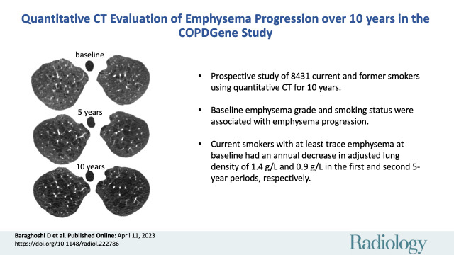
Background Long-term studies of chronic obstructive pulmonary disease (COPD) can evaluate emphysema progression. Adjustment for differences in equipment and scanning protocols of individual CT examinations have not been studied extensively. Purpose To evaluate emphysema progression in current and former smokers in the COPDGene cohort over three imaging points obtained at 5-year intervals accounting for individual CT parameters. Materials and Methods Current and former cigarette smokers enrolled between 2008 and 2011 from the COPDGene study were prospectively followed for 10 years between 2008 and 2020. Extent of emphysema as adjusted lung density (ALD) from quantitative CT was measured at baseline and at 5- and 10-year follow-up. Linear mixed models adjusted for CT technical characteristics were constructed to evaluate emphysema progression. Mean annual changes in ALD over consecutive 5-year study periods were estimated by smoking status and baseline emphysema. Results Of 8431 participants at baseline (mean age, 60 years ± 9 [SD]; 3905 female participants), 4913 were at 5-year follow-up and 1544 participants were at 10-year follow-up. There were 4134 (49%) participants who were current smokers, and 4449 (53%) participants had more than trace emphysema at baseline. Current smokers with more than trace emphysema showed the largest decline in ALD, with mean annual decreases of 1.4 g/L (95% CI: 1.2, 1.5) in the first 5 years and 0.9 g/L (95% CI: 0.7, 1.2) in the second 5 years. Accounting for CT noise, field of view, and scanner model improved model fit for estimation of emphysema progression ( < .001 by likelihood ratio test). Conclusion Evaluation at CT of emphysema progression in the COPDGene study showed that, during the span of 10 years, participants with pre-existing emphysema who continued smoking had the largest decline in ALD. Adjusting for CT equipment and protocol factors improved these longitudinal estimates. Clinical trial registration no. NCT00608764 © RSNA, 2023 See the editorial by Parraga and Kirby in this issue.
长期的慢性阻塞性肺疾病(COPD)研究可以评估肺气肿的进展。但是,针对个体 CT 检查的设备和扫描方案的差异,尚未进行广泛的调整。目的:在 COPDGene 队列中,通过在 5 年的时间间隔内进行 3 次 CT 检查,评估当前吸烟者和曾经吸烟者的肺气肿进展情况,并考虑到个体 CT 参数。材料与方法:本研究前瞻性地随访了 COPDGene 研究中于 2008 年至 2011 年期间入组的当前吸烟者和曾经吸烟者,随访时间为 2008 年至 2020 年共 10 年。在基线和 5 年及 10 年的随访时,使用定量 CT 测量调整后的肺密度(ALD)。构建线性混合模型,以调整 CT 技术特征,来评估肺气肿的进展情况。通过吸烟状态和基线肺气肿情况,估计连续 5 年研究期间 ALD 的平均年变化。结果:在 8431 名基线参与者中(平均年龄为 60 岁±9[标准差];3905 名女性参与者),有 4913 名在 5 年随访时,1544 名在 10 年随访时完成了检查。其中 4134 名(49%)为当前吸烟者,4449 名(53%)基线时存在微量肺气肿。有微量肺气肿的当前吸烟者的 ALD 下降幅度最大,在最初的 5 年中,ALD 的平均年下降率为 1.4 g/L(95%CI:1.2,1.5),在随后的 5 年中,ALD 的平均年下降率为 0.9 g/L(95%CI:0.7,1.2)。考虑到 CT 噪声、视野和扫描仪型号,改善了对肺气肿进展的模型拟合(似然比检验,P<.001)。结论:在 COPDGene 研究中,对 CT 评估的肺气肿进展情况进行评估后发现,在 10 年的时间跨度内,持续吸烟且存在肺气肿的参与者的 ALD 下降幅度最大。调整 CT 设备和方案因素后,可提高这些纵向估计值。临床试验注册号:NCT00608764