Department of Medicine and Surgery, University of Parma, 43126 Parma, Italy.
Department of Occupational and Environmental Medicine, Epidemiology and Hygiene, Italian Workers' Compensation Authority-INAIL, 00078 Rome, Italy.
Int J Mol Sci. 2023 Apr 1;24(7):6595. doi: 10.3390/ijms24076595.
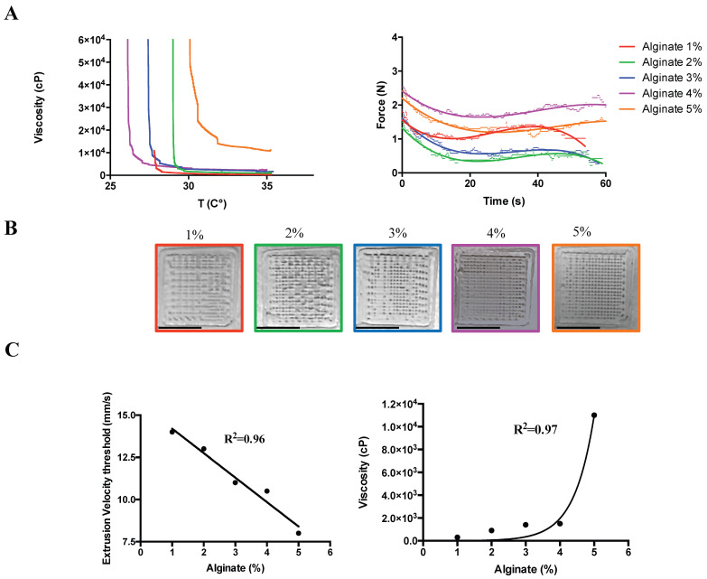
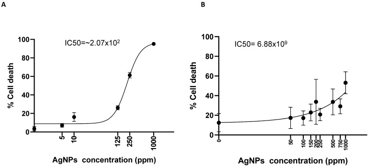
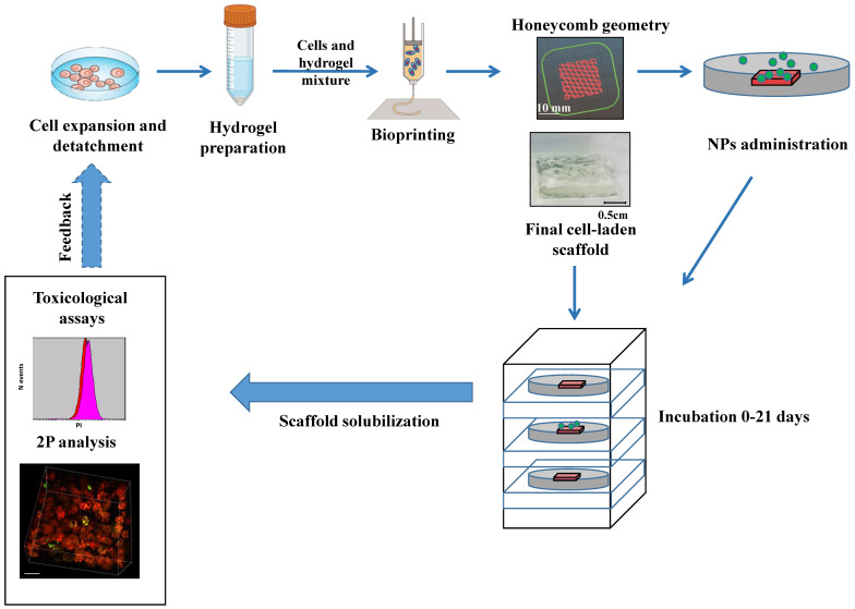
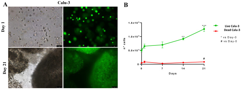
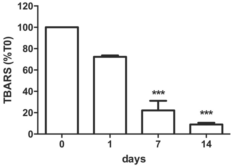
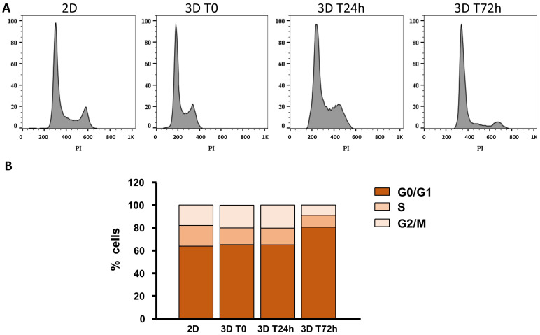
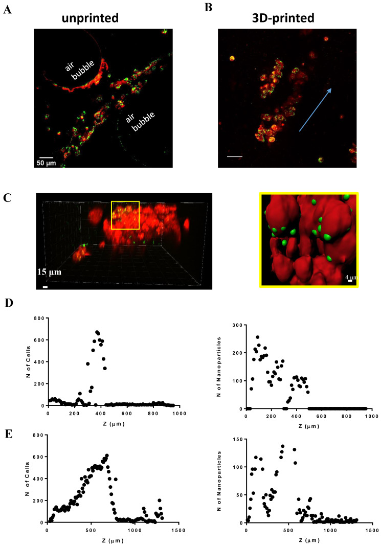
The toxicity of nanoparticles absorbed through contact or inhalation is one of the major concerns for public health. It is mandatory to continually evaluate the toxicity of nanomaterials. In vitro nanotoxicological studies are conventionally limited by the two dimensions. Although 3D bioprinting has been recently adopted for three-dimensional culture in the context of drug release and tissue regeneration, little is known regarding its use for nanotoxicology investigation. Therefore, aiming to simulate the exposure of lung cells to nanoparticles, we developed organoid-based scaffolds for long-term studies in immortalized cell lines. We printed the viscous cell-laden material via a customized 3D bioprinter and subsequently exposed the scaffold to either 40 nm latex-fluorescent or 11-14 nm silver nanoparticles. The number of cells significantly increased on the 14th day in the 3D environment, from 5 × 10 to 1.27 × 10, showing a 91% lipid peroxidation reduction over time and minimal cell death observed throughout 21 days. Administered fluorescent nanoparticles can diffuse throughout the 3D-printed scaffolds while this was not the case for the unprinted ones. A significant increment in cell viability from 3D vs. 2D cultures exposed to silver nanoparticles has been demonstrated. This shows toxicology responses that recapitulate in vivo experiments, such as inhaled silver nanoparticles. The results open a new perspective in 3D protocols for nanotoxicology investigation supporting 3Rs.
纳米颗粒通过接触或吸入而被吸收所产生的毒性是公众健康的主要关注点之一。因此,必须不断评估纳米材料的毒性。传统的体外纳米毒理学研究通常受到二维的限制。尽管 3D 生物打印最近已被用于药物释放和组织再生的三维培养,但对于其在纳米毒理学研究中的应用知之甚少。因此,为了模拟肺细胞暴露于纳米颗粒的情况,我们开发了基于类器官的支架,用于在永生化细胞系中进行长期研究。我们通过定制的 3D 生物打印机打印粘性细胞负载材料,然后将支架暴露于 40nm 乳胶荧光或 11-14nm 银纳米颗粒下。在 3D 环境中,细胞数量在第 14 天显著增加,从 5×10 增加到 1.27×10,显示出随着时间的推移脂质过氧化减少 91%,整个 21 天内观察到的细胞死亡最小。可扩散进入 3D 打印支架的荧光纳米颗粒而未打印的纳米颗粒则无法扩散进入。暴露于银纳米颗粒的 3D 培养物与 2D 培养物相比,细胞活力显著增加。这表明毒性反应与体内实验(如吸入的银纳米颗粒)相似。这些结果为支持 3R 的纳米毒理学研究的 3D 方案开辟了新的视角。