Department of Biology, University of Padova, Via Ugo Bassi 58B, 35131 Padova, Italy.
Department of Medicine, University of Udine, Piazzale Kolbe, 33100 Udine, Italy.
Cells. 2023 Apr 5;12(7):1089. doi: 10.3390/cells12071089.
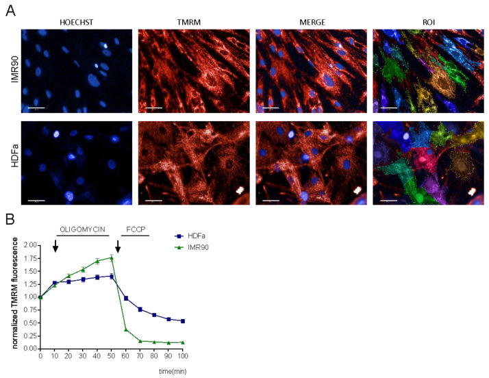
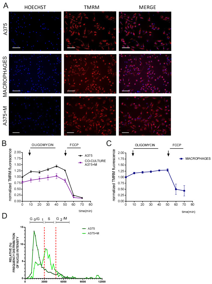
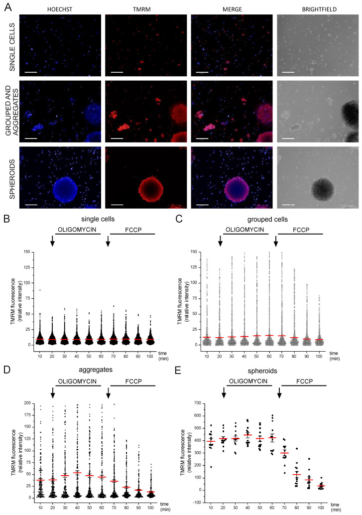
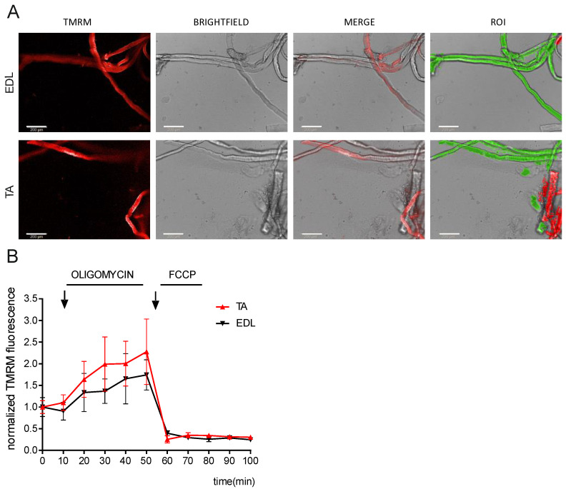
Recent proteomic, metabolomic, and transcriptomic studies have highlighted a connection between changes in mitochondria physiology and cellular pathophysiological mechanisms. Secondary assays to assess the function of these organelles appear fundamental to validate these -omics findings. Although mitochondrial membrane potential is widely recognized as an indicator of mitochondrial activity, high-content imaging-based approaches coupled to multiparametric to measure it have not been established yet. In this paper, we describe a methodology for the unbiased high-throughput quantification of mitochondrial membrane potential in vitro, which is suitable for 2D to 3D models. We successfully used our method to analyze mitochondrial membrane potential in monolayers of human fibroblasts, neural stem cells, spheroids, and isolated muscle fibers. Moreover, by combining automated image analysis and machine learning, we were able to discriminate melanoma cells from macrophages in co-culture and to analyze the subpopulations separately. Our data demonstrated that our method is a widely applicable strategy for large-scale profiling of mitochondrial activity.
最近的蛋白质组学、代谢组学和转录组学研究强调了线粒体生理学变化与细胞病理生理机制之间的联系。评估这些细胞器功能的辅助检测似乎对于验证这些组学发现至关重要。尽管线粒体膜电位被广泛认为是线粒体活性的指标,但尚未建立基于高内涵成像的方法来结合多参数进行测量。在本文中,我们描述了一种用于体外高通量量化线粒体膜电位的无偏方法,该方法适用于 2D 至 3D 模型。我们成功地使用我们的方法分析了单层人成纤维细胞、神经干细胞、球体和分离的肌肉纤维中的线粒体膜电位。此外,通过将自动图像分析和机器学习相结合,我们能够区分共培养中的黑色素瘤细胞和巨噬细胞,并分别对亚群进行分析。我们的数据表明,我们的方法是一种广泛适用于大规模线粒体活性分析的策略。