• 文献检索
  • 文档翻译
  • 深度研究
  • 学术资讯
  • Suppr Zotero 插件Zotero 插件
  • 邀请有礼
  • 套餐&价格
  • 历史记录
应用&插件
Suppr Zotero 插件Zotero 插件浏览器插件Mac 客户端Windows 客户端微信小程序
定价
高级版会员购买积分包购买API积分包
服务
文献检索文档翻译深度研究API 文档MCP 服务
关于我们
关于 Suppr公司介绍联系我们用户协议隐私条款
关注我们

Suppr 超能文献

核心技术专利:CN118964589B侵权必究
粤ICP备2023148730 号-1Suppr @ 2026

文献检索

告别复杂PubMed语法,用中文像聊天一样搜索,搜遍4000万医学文献。AI智能推荐,让科研检索更轻松。

立即免费搜索

文件翻译

保留排版,准确专业,支持PDF/Word/PPT等文件格式,支持 12+语言互译。

免费翻译文档

深度研究

AI帮你快速写综述,25分钟生成高质量综述,智能提取关键信息,辅助科研写作。

立即免费体验

软组织肉瘤放疗反应监测的多参数磁共振成像:一种组织学与 MRI 配准算法。

Multiparametric magnetic resonance imaging for radiation therapy response monitoring in soft tissue sarcomas: a histology and MRI co-registration algorithm.

机构信息

Department of Diagnostic and Interventional Radiology, University Medical Center Freiburg, Faculty of Medicine, University of Freiburg, Germany.

Berta-Ottenstein-Programme, University of Freiburg, Faculty of Medicine, Freiburg, Germany.

出版信息

Theranostics. 2023 Mar 5;13(5):1594-1606. doi: 10.7150/thno.81938. eCollection 2023.

DOI:10.7150/thno.81938
PMID:37056570
原文链接:https://pmc.ncbi.nlm.nih.gov/articles/PMC10086213/
Abstract

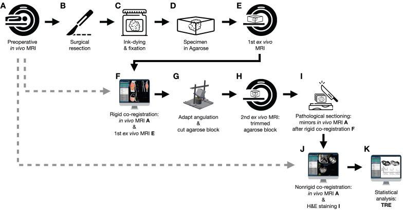
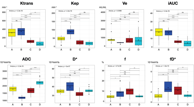
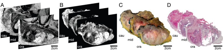
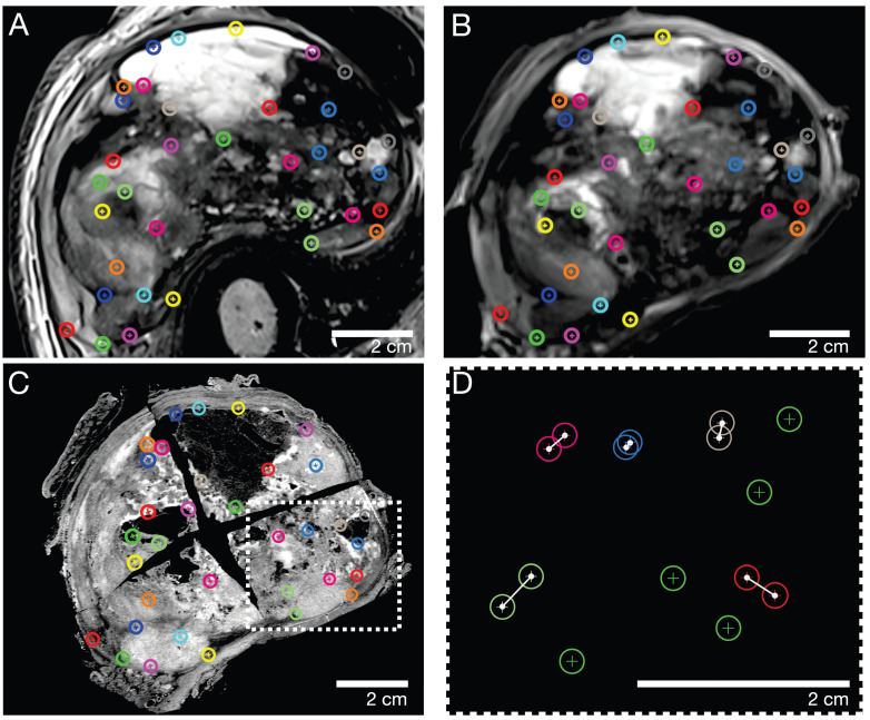
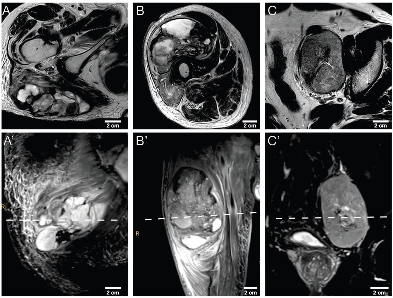
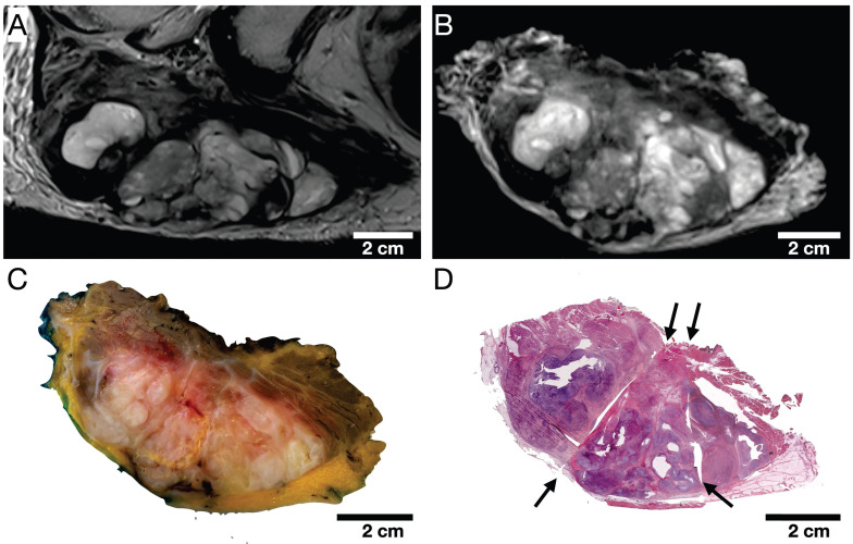
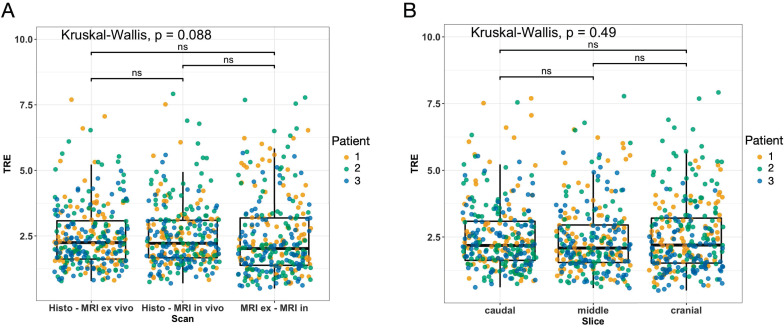
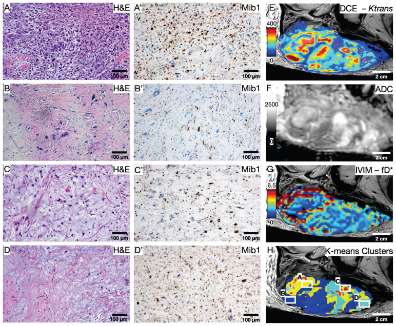
To establish a spatially exact co-registration procedure between multiparametric magnetic resonance imaging (mpMRI) and (immuno)histopathology of soft tissue sarcomas (STS) to identify imaging parameters that reflect radiation therapy response of STS. The mpMRI-Protocol included diffusion-weighted (DWI), intravoxel-incoherent motion (IVIM), and dynamic contrast-enhancing (DCE) imaging. The resection specimen was embedded in 6.5% agarose after initial fixation in formalin. To ensure identical alignment of histopathological sectioning and imaging, an MRI scan of the specimen was rigidly co-registered with the mpMRI. The deviating angulation of the specimen to the location of the tumor was determined. The agarose block was trimmed accordingly. A second MRI in a dedicated localizer with a 4 mm grid was performed, which was matched to a custom-built sectioning machine. Microtomy sections were stained with hematoxylin and eosin. Immunohistochemical staining was performed with anti-ALDH1A1 antibodies as a radioresistance and anti-MIB1 antibodies as a proliferation marker. Fusion of the digitized microtomy sections with the mpMRI was accomplished through nonrigid co-registration to the mpMRI. Co-registration accuracy was qualitatively assessed by visual assessment and quantitatively evaluated by computing target registration errors (TRE). The study sample comprised nine tumor sections from three STS patients. Visual assessment after nonrigid co-registration showed a strong morphological correlation of the histopathological specimens with MRI and mpMRI after neoadjuvant radiation therapy. Quantitative assessment of the co-registration procedure using TRE analysis of different pairs of pathology and MRI sections revealed highly accurate structural alignment, with a total median TRE of 2.25 mm (histology - MRI), 2.22 mm (histology - mpMRI), and 2.02 mm ( MRI - mpMRI). There was no significant difference between TREs of the different pairs of sections or caudal, middle, and cranial tumor parts, respectively. Our initial results show a promising approach to obtaining accurate co-registration between histopathology and MRI for STS. In a larger cohort of patients, the method established here will enable the prospective identification and validation of imaging biomarkers for radiation therapy response prediction and monitoring in STS patients via precise molecular and cellular correlation.

摘要

为了在软组织肉瘤(STS)的多参数磁共振成像(mpMRI)和(免疫)组织病理学之间建立空间精确的配准程序,以确定反映 STS 放射治疗反应的成像参数。mpMRI 方案包括扩散加权成像(DWI)、体素内不相干运动(IVIM)和动态对比增强成像(DCE)。切除标本在福尔马林初始固定后嵌入 6.5%琼脂糖中。为了确保组织病理学切片和成像的精确对齐,对标本进行刚性配准与 mpMRI。确定标本与肿瘤位置的偏离角度。相应地修剪琼脂糖块。在专用定位器中进行第二次具有 4 毫米网格的 MRI,与定制的切片机匹配。对苏木精和伊红染色的微切片进行染色。使用抗 ALDH1A1 抗体作为放射抗性和抗 MIB1 抗体作为增殖标志物进行免疫组织化学染色。通过与 mpMRI 的非刚性配准来实现数字化微切片与 mpMRI 的融合。通过视觉评估定性评估配准准确性,并通过计算目标配准误差(TRE)定量评估配准准确性。该研究样本包括来自三名 STS 患者的九个肿瘤切片。在新辅助放射治疗后,非刚性配准后的数字评估显示,组织病理学标本与 MRI 和 mpMRI 之间具有很强的形态相关性。使用不同的病理学和 MRI 切片对配准过程进行 TRE 分析的定量评估显示,结构对准高度准确,总中位数 TRE 为 2.25 毫米(组织学 - MRI)、2.22 毫米(组织学 - mpMRI)和 2.02 毫米(MRI - mpMRI)。不同切片对之间或尾、中、颅肿瘤部分的 TRE 之间没有显著差异。我们的初步结果表明,这是一种在 STS 中获得组织病理学和 MRI 之间精确配准的有前途的方法。在更大的患者队列中,这里建立的方法将能够通过精确的分子和细胞相关性,前瞻性地识别和验证用于预测和监测 STS 患者放射治疗反应的成像生物标志物。

https://cdn.ncbi.nlm.nih.gov/pmc/blobs/5b28/10086213/b2b49cad7626/thnov13p1594g008.jpg
https://cdn.ncbi.nlm.nih.gov/pmc/blobs/5b28/10086213/10192c14b7f7/thnov13p1594g001.jpg
https://cdn.ncbi.nlm.nih.gov/pmc/blobs/5b28/10086213/f8eb342985d9/thnov13p1594g002.jpg
https://cdn.ncbi.nlm.nih.gov/pmc/blobs/5b28/10086213/372d92c0f68d/thnov13p1594g003.jpg
https://cdn.ncbi.nlm.nih.gov/pmc/blobs/5b28/10086213/9cfffe09da0e/thnov13p1594g004.jpg
https://cdn.ncbi.nlm.nih.gov/pmc/blobs/5b28/10086213/0d592ea4c815/thnov13p1594g005.jpg
https://cdn.ncbi.nlm.nih.gov/pmc/blobs/5b28/10086213/95a887f5578a/thnov13p1594g006.jpg
https://cdn.ncbi.nlm.nih.gov/pmc/blobs/5b28/10086213/ceffdc751aec/thnov13p1594g007.jpg
https://cdn.ncbi.nlm.nih.gov/pmc/blobs/5b28/10086213/b2b49cad7626/thnov13p1594g008.jpg

相似文献

1
Multiparametric magnetic resonance imaging for radiation therapy response monitoring in soft tissue sarcomas: a histology and MRI co-registration algorithm.软组织肉瘤放疗反应监测的多参数磁共振成像:一种组织学与 MRI 配准算法。
Theranostics. 2023 Mar 5;13(5):1594-1606. doi: 10.7150/thno.81938. eCollection 2023.
2
Non-invasive monitoring of neoadjuvant radiation therapy response in soft tissue sarcomas by multiparametric MRI and quantification of circulating tumor DNA-A study protocol.多参数 MRI 无创监测软组织肉瘤新辅助放疗反应及循环肿瘤 DNA 定量:一项研究方案。
PLoS One. 2023 Nov 1;18(11):e0285580. doi: 10.1371/journal.pone.0285580. eCollection 2023.
3
Development of a registration framework to validate MRI with histology for prostate focal therapy.开发一个用于前列腺聚焦治疗的磁共振成像(MRI)与组织学验证的注册框架。
Med Phys. 2015 Dec;42(12):7078-89. doi: 10.1118/1.4935343.
4
Voxel-wise correlation of positron emission tomography/computed tomography with multiparametric magnetic resonance imaging and histology of the prostate using a sophisticated registration framework.利用复杂的配准框架,对前列腺进行正电子发射断层扫描/计算机断层扫描与多参数磁共振成像和组织学的体素相关性研究。
BJU Int. 2019 Jun;123(6):1020-1030. doi: 10.1111/bju.14648. Epub 2019 Jan 28.
5
Multi-scale statistical deformation based co-registration of prostate MRI and post-surgical whole mount histopathology.基于多尺度统计变形的前列腺MRI与术后全层组织病理学图像配准
Med Phys. 2024 Apr;51(4):2549-2562. doi: 10.1002/mp.16753. Epub 2023 Sep 24.
6
Preliminary experience with a novel method of three-dimensional co-registration of prostate cancer digital histology and in vivo multiparametric MRI.前列腺癌数字组织病理学与体内多参数 MRI 三维配准新方法的初步经验。
Clin Radiol. 2013 Dec;68(12):e652-8. doi: 10.1016/j.crad.2013.07.010. Epub 2013 Aug 28.
7
Multiparametric Magnetic Resonance Imaging as a Noninvasive Assessment of Tumor Response to Neoadjuvant Pembrolizumab in Muscle-invasive Bladder Cancer: Preliminary Findings from the PURE-01 Study.多参数磁共振成像作为新辅助帕博利珠单抗治疗肌层浸润性膀胱癌肿瘤反应的无创评估:来自 PURE-01 研究的初步发现。
Eur Urol. 2020 May;77(5):636-643. doi: 10.1016/j.eururo.2019.12.016. Epub 2019 Dec 25.
8
Habitat escalated adaptive therapy (HEAT): a phase 2 trial utilizing radiomic habitat-directed and genomic-adjusted radiation dose (GARD) optimization for high-grade soft tissue sarcoma.生境升级适应性治疗(HEAT):一项利用放射组学生态位导向和基因组调整的辐射剂量(GARD)优化治疗高级软组织肉瘤的 2 期试验。
BMC Cancer. 2024 Apr 9;24(1):437. doi: 10.1186/s12885-024-12151-7.
9
Impact of Machine Learning With Multiparametric Magnetic Resonance Imaging of the Breast for Early Prediction of Response to Neoadjuvant Chemotherapy and Survival Outcomes in Breast Cancer Patients.机器学习联合乳腺多参数磁共振成像对乳腺癌新辅助化疗早期疗效及生存预后评估的影响。
Invest Radiol. 2019 Feb;54(2):110-117. doi: 10.1097/RLI.0000000000000518.
10
Multiparametric MRI model with dynamic contrast-enhanced and diffusion-weighted imaging enables breast cancer diagnosis with high accuracy.多参数 MRI 模型结合动态对比增强和弥散加权成像可实现高准确率的乳腺癌诊断。
J Magn Reson Imaging. 2019 Mar;49(3):864-874. doi: 10.1002/jmri.26285. Epub 2018 Oct 30.

引用本文的文献

1
Research on application of multiparametric MRI to predict FNCLCC grading and ki67 expression in soft tissue sarcoma biopsy pathology: Based on a CT-MRI fusion image registration method.基于CT-MRI融合图像配准方法的多参数MRI在软组织肉瘤活检病理中预测FNCLCC分级及Ki67表达的应用研究
Front Oncol. 2025 Aug 8;15:1606942. doi: 10.3389/fonc.2025.1606942. eCollection 2025.
2
Biomarker-driven molecular imaging probes in radiotherapy.基于生物标志物的放射治疗分子影像探针。
Theranostics. 2024 Jul 2;14(10):4127-4146. doi: 10.7150/thno.97768. eCollection 2024.
3
Non-invasive monitoring of neoadjuvant radiation therapy response in soft tissue sarcomas by multiparametric MRI and quantification of circulating tumor DNA-A study protocol.

本文引用的文献

1
Soft tissue sarcoma: correlation of dynamic contrast-enhanced magnetic resonance imaging features with HIF-1α expression and patient outcomes.软组织肉瘤:动态对比增强磁共振成像特征与缺氧诱导因子-1α表达及患者预后的相关性
Quant Imaging Med Surg. 2022 Oct;12(10):4823-4836. doi: 10.21037/qims-22-75.
2
Registration of histopathology to magnetic resonance imaging of prostate cancer.前列腺癌组织病理学与磁共振成像的配准
Phys Imaging Radiat Oncol. 2021 Apr 12;18:19-25. doi: 10.1016/j.phro.2021.03.004. eCollection 2021 Apr.
3
Perfusion-driven Intravoxel Incoherent Motion (IVIM) MRI in Oncology: Applications, Challenges, and Future Trends.
多参数 MRI 无创监测软组织肉瘤新辅助放疗反应及循环肿瘤 DNA 定量:一项研究方案。
PLoS One. 2023 Nov 1;18(11):e0285580. doi: 10.1371/journal.pone.0285580. eCollection 2023.
4
[Ga]Ga-PSMA and [Ga]Ga-RM2 PET/MRI vs. Histopathological Images in Prostate Cancer: A New Workflow for Spatial Co-Registration.[镓]镓-PSMA和[镓]镓-RM2正电子发射断层显像/磁共振成像与前列腺癌组织病理学图像的对比:一种空间配准的新工作流程
Bioengineering (Basel). 2023 Aug 11;10(8):953. doi: 10.3390/bioengineering10080953.
灌注驱动的体素内不相干运动(IVIM)磁共振成像在肿瘤学中的应用、挑战与未来趋势。
Magn Reson Med Sci. 2021 Jun 1;20(2):125-138. doi: 10.2463/mrms.rev.2019-0124. Epub 2020 Jun 15.
4
Multidisciplinary treatment of soft tissue sarcomas: An update.软组织肉瘤的多学科治疗:最新进展
World J Clin Oncol. 2020 Apr 24;11(4):180-189. doi: 10.5306/wjco.v11.i4.180.
5
Soft tissue sarcoma: DWI and DCE-MRI parameters correlate with Ki-67 labeling index.软组织肉瘤:DWI 和 DCE-MRI 参数与 Ki-67 标记指数相关。
Eur Radiol. 2020 Feb;30(2):914-924. doi: 10.1007/s00330-019-06445-9. Epub 2019 Oct 18.
6
Incidence, distribution of histological subtypes and primary sites of soft tissue sarcoma in China.中国软组织肉瘤的发病率、组织学亚型分布及原发部位
Cancer Biol Med. 2019 Aug;16(3):565-574. doi: 10.20892/j.issn.2095-3941.2019.0041.
7
Multiparametric MRI and Coregistered Histology Identify Tumor Habitats in Breast Cancer Mouse Models.多参数 MRI 与配准组织学鉴定乳腺癌小鼠模型中的肿瘤微环境。
Cancer Res. 2019 Aug 1;79(15):3952-3964. doi: 10.1158/0008-5472.CAN-19-0213. Epub 2019 Jun 11.
8
Utility of Multi-Parametric Quantitative Magnetic Resonance Imaging for Characterization and Radiotherapy Response Assessment in Soft-Tissue Sarcomas and Correlation With Histopathology.多参数定量磁共振成像在软组织肉瘤特征描述及放疗反应评估中的应用及其与组织病理学的相关性
Front Oncol. 2019 Apr 25;9:280. doi: 10.3389/fonc.2019.00280. eCollection 2019.
9
Soft-Tissue Sarcomas: Assessment of MRI Features Correlating with Histologic Grade and Patient Outcome.软组织肉瘤:与组织学分级和患者预后相关的 MRI 特征评估。
Radiology. 2019 Jun;291(3):710-721. doi: 10.1148/radiol.2019181659. Epub 2019 Apr 9.
10
A system using patient-specific 3D-printed molds to spatially align in vivo MRI with ex vivo MRI and whole-mount histopathology for prostate cancer research.一种使用患者特异性 3D 打印模具的系统,用于将体内 MRI 与体外 MRI 和全组织病理切片进行空间配准,以进行前列腺癌研究。
J Magn Reson Imaging. 2019 Jan;49(1):270-279. doi: 10.1002/jmri.26189. Epub 2018 Aug 2.