Sandgren Kristina, Nilsson Erik, Keeratijarut Lindberg Angsana, Strandberg Sara, Blomqvist Lennart, Bergh Anders, Friedrich Bengt, Axelsson Jan, Ögren Margareta, Ögren Mattias, Widmark Anders, Thellenberg Karlsson Camilla, Söderkvist Karin, Riklund Katrine, Jonsson Joakim, Nyholm Tufve
Department of Radiation Sciences, Radiophysics, Umea University, Sweden.
Department of Radiation Sciences, Diagnostic Radiology, Umea University, Sweden.
Phys Imaging Radiat Oncol. 2021 Apr 12;18:19-25. doi: 10.1016/j.phro.2021.03.004. eCollection 2021 Apr.
The diagnostic accuracy of new imaging techniques requires validation, preferably by histopathological verification. The aim of this study was to develop and present a registration procedure between histopathology and magnetic resonance imaging (MRI) of the prostate, to estimate its uncertainty and to evaluate the benefit of adding a contour-correcting registration.
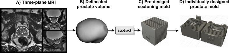
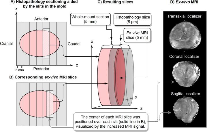
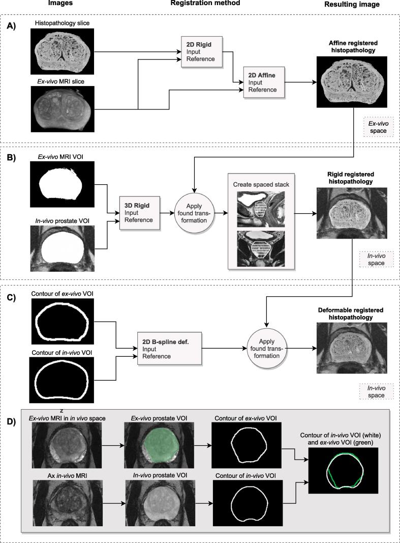
For twenty-five prostate cancer patients, planned for radical prostatectomy, a 3D-printed prostate mold based on MRI was created and an MRI of the specimen, placed inside the mold, was performed. Each histopathology slice was registered to its corresponding MRI slice using a 2D-affine registration. The MRI was rigidly registered to the MRI and the resulting transform was applied to the histopathology stack. A 2D deformable registration was used to correct for specimen distortion concerning the specimen's fit inside the mold. We estimated the spatial uncertainty by comparing positions of landmarks in the MRI and the corresponding registered histopathology stack.
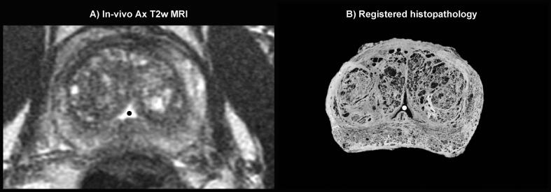
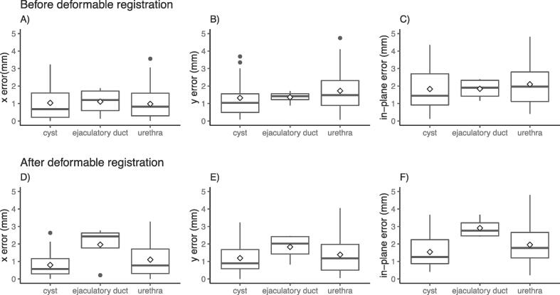
Eighty-four landmarks were identified, located in the urethra (62%), prostatic cysts (33%), and the ejaculatory ducts (5%). The median number of landmarks was 3 per patient. We showed a median in-plane error of 1.8 mm before and 1.7 mm after the contour-correcting deformable registration. In patients with extraprostatic margins, the median in-plane error improved from 2.1 mm to 1.8 mm after the contour-correcting deformable registration.
Our registration procedure accurately registers histopathology to MRI, with low uncertainty. The contour-correcting registration was beneficial in patients with extraprostatic surgical margins.
新成像技术的诊断准确性需要验证,最好通过组织病理学验证。本研究的目的是开发并展示一种前列腺组织病理学与磁共振成像(MRI)之间的配准程序,估计其不确定性,并评估添加轮廓校正配准的益处。
对于计划进行根治性前列腺切除术的25例前列腺癌患者,制作了基于MRI的3D打印前列腺模具,并对置于模具内的标本进行了MRI检查。使用二维仿射配准将每个组织病理学切片与其对应的MRI切片进行配准。将MRI与MRI进行刚性配准,并将所得变换应用于组织病理学图像堆栈。使用二维可变形配准来校正标本在模具内拟合时的变形。我们通过比较MRI和相应配准组织病理学图像堆栈中标记点的位置来估计空间不确定性。
共识别出84个标记点,位于尿道(62%)、前列腺囊肿(33%)和射精管(5%)。每位患者标记点的中位数为3个。我们发现,在进行轮廓校正可变形配准之前,平面内误差中位数为1.8毫米,之后为1.7毫米。对于有前列腺外切缘的患者,轮廓校正可变形配准后,平面内误差中位数从2.1毫米改善至1.8毫米。
我们的配准程序能准确地将组织病理学与MRI配准,不确定性较低。轮廓校正配准对有前列腺外手术切缘的患者有益。