Martínez Martín, Ariz Mikel, Alvarez Ignacio, Castellanos Gabriel, Aguilar Miquel, Hernández-Vara Jorge, Caballol Núria, Garrido Alicia, Bayés Àngels, Vilas Dolores, Marti Maria Jose, Pastor Pau, de Solórzano Carlos Ortiz, Pastor Maria A
Neuroimaging Laboratory, University of Navarra, School of Medicine, Pamplona, Spain.
School of Education and Psychology, University of Navarra, Pamplona, Spain.
NPJ Parkinsons Dis. 2023 Apr 15;9(1):62. doi: 10.1038/s41531-023-00503-2.
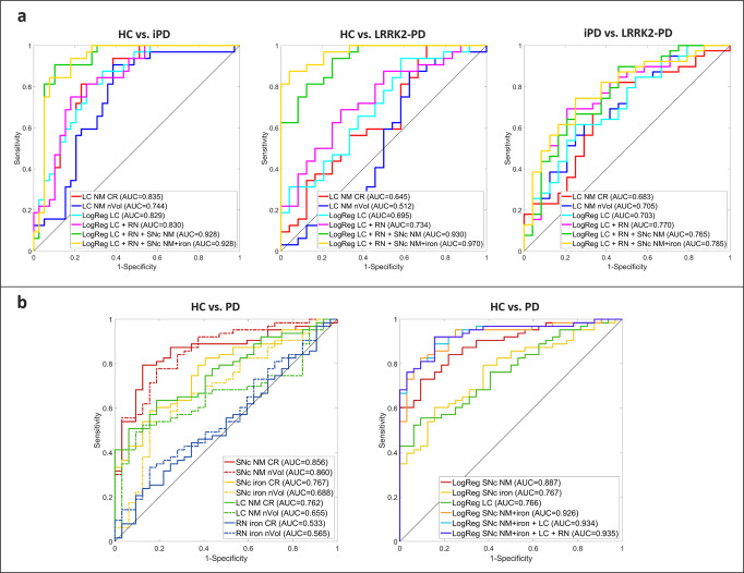
Neuromelanin (NM) loss in substantia nigra pars compacta (SNc) and locus coeruleus (LC) reflects neuronal death in Parkinson's disease (PD). Since genetically-determined PD shows varied clinical expressivity, we wanted to accurately quantify and locate brainstem NM and iron, to discover whether specific MRI patterns are linked to Leucine-rich repeat kinase 2 G2019S PD (LRRK2-PD) or idiopathic Parkinson's disease (iPD). A 3D automated MRI atlas-based segmentation pipeline (3D-ABSP) for NM/iron-sensitive MRI images topographically characterized the SNc, LC, and red nucleus (RN) neuronal loss and calculated NM/iron contrast ratio (CR) and normalized volume (nVol). Left-side NM nVol was larger in all groups. PD had lower NM CR and nVol in ventral-caudal SNc, whereas iron increased in lateral, medial-rostral, and caudal SNc. The SNc NM CR reduction was associated with psychiatric symptoms. LC CR and nVol discriminated better among subgroups: LRRK2-PD had similar LC NM CR and nVol as that of controls, and larger LC NM nVol and RN iron CR than iPD. PD showed higher iron SNc nVol than controls, especially among LRRK2-PD. ROC analyses showed an AUC > 0.92 for most pairwise subgroup comparisons, with SNc NM being the best discriminator between HC and PD. NM measures maintained their discriminator power considering the subgroup of PD patients with less than 5 years of disease duration. The SNc iron CR and nVol increase was associated with longer disease duration in PD patients. The 3D-ABSP sensitively identified NM and iron MRI patterns strongly correlated with phenotypic PD features.
黑质致密部(SNc)和蓝斑(LC)中神经黑色素(NM)的缺失反映了帕金森病(PD)中的神经元死亡。由于基因决定的PD表现出不同的临床表型,我们希望准确量化和定位脑干中的NM和铁,以发现特定的MRI模式是否与富含亮氨酸重复激酶2 G2019S型PD(LRRK2-PD)或特发性帕金森病(iPD)相关。一种基于3D自动MRI图谱的分割管道(3D-ABSP),用于对NM/铁敏感的MRI图像,从地形学上表征了SNc、LC和红核(RN)的神经元损失,并计算了NM/铁对比度(CR)和标准化体积(nVol)。所有组中左侧NM的nVol均较大。PD患者腹侧尾侧SNc的NM CR和nVol较低,而外侧、内侧 Rostral 和尾侧SNc的铁含量增加。SNc中NM CR的降低与精神症状有关。LC的CR和nVol在亚组之间的区分更好:LRRK2-PD的LC NM CR和nVol与对照组相似,且LC NM nVol和RN铁CR比iPD更大。PD患者SNc的铁nVol高于对照组,尤其是在LRRK2-PD患者中。ROC分析显示,大多数两两亚组比较的AUC>0.92,SNc中的NM是健康对照(HC)和PD之间的最佳区分指标。考虑到病程小于5年的PD患者亚组,NM测量值保持了其区分能力。PD患者中SNc铁CR和nVol的增加与病程延长有关。3D-ABSP敏感地识别出与PD表型特征密切相关的NM和铁MRI模式。