Department of Paediatric Infectious Diseases, Amsterdam UMC Location University of Amsterdam, Amsterdam, The Netherlands.
Department Medical Microbiology, OrganoVIR Labs, Amsterdam UMC Location University of Amsterdam, Amsterdam, the Netherlands.
Stem Cell Res Ther. 2023 Apr 15;14(1):87. doi: 10.1186/s13287-023-03302-x.
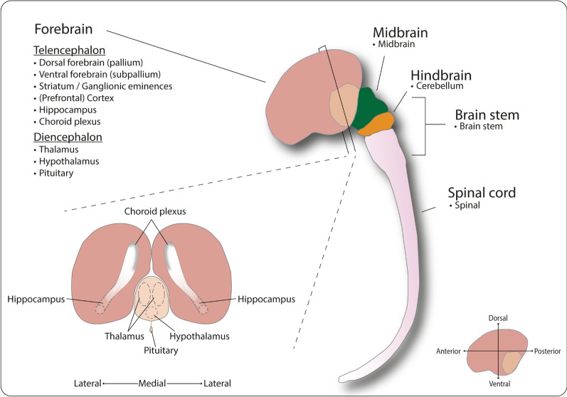
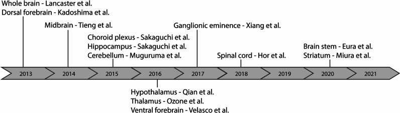
The first human brain organoid protocol was presented in the beginning of the previous decade, and since then, the field witnessed the development of many new brain region-specific models, and subsequent protocol adaptations and modifications. The vast amount of data available on brain organoid technology may be overwhelming for scientists new to the field and consequently decrease its accessibility. Here, we aimed at providing a practical guide for new researchers in the field by systematically reviewing human brain organoid publications.
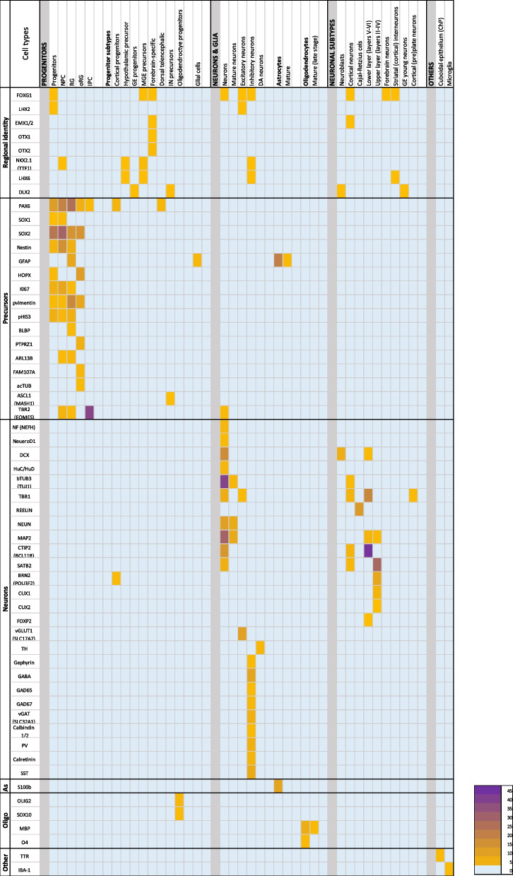
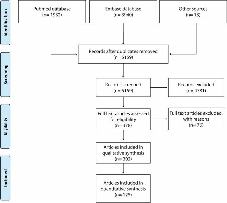
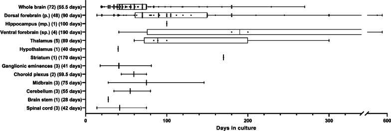
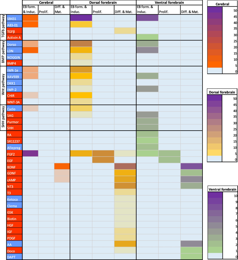
Articles published between 2010 and 2020 were selected and categorised for brain organoid applications. Those describing neurodevelopmental studies or protocols for novel organoid models were further analysed for culture duration of the brain organoids, protocol comparisons of key aspects of organoid generation, and performed functional characterisation assays. We then summarised the approaches taken for different models and analysed the application of small molecules and growth factors used to achieve organoid regionalisation. Finally, we analysed articles for organoid cell type compositions, the reported time points per cell type, and for immunofluorescence markers used to characterise different cell types.
Calcium imaging and patch clamp analysis were the most frequently used neuronal activity assays in brain organoids. Neural activity was shown in all analysed models, yet network activity was age, model, and assay dependent. Induction of dorsal forebrain organoids was primarily achieved through combined (dual) SMAD and Wnt signalling inhibition. Ventral forebrain organoid induction was performed with dual SMAD and Wnt signalling inhibition, together with additional activation of the Shh pathway. Cerebral organoids and dorsal forebrain model presented the most cell types between days 35 and 60. At 84 days, dorsal forebrain organoids contain astrocytes and potentially oligodendrocytes. Immunofluorescence analysis showed cell type-specific application of non-exclusive markers for multiple cell types.
We provide an easily accessible overview of human brain organoid cultures, which may help those working with brain organoids to define their choice of model, culture time, functional assay, differentiation, and characterisation strategies.
第一个人类大脑类器官的方案在十年前提出,从那时起,该领域见证了许多新的脑区特异性模型的发展,以及随后的方案适应和修改。对于该领域的新手科学家来说,大量可获得的大脑类器官技术数据可能令人不知所措,从而降低其可及性。在这里,我们旨在通过系统地审查人类大脑类器官的文献,为该领域的新研究人员提供实用指南。
选择并分类了 2010 年至 2020 年期间发表的文章,用于脑类器官应用。那些描述神经发育研究或新型类器官模型方案的文章,进一步分析了脑类器官的培养时间、类器官生成关键方面的方案比较,以及进行功能特征分析的方法。然后,我们总结了不同模型采用的方法,并分析了用于实现类器官区域化的小分子和生长因子的应用。最后,我们分析了文章中的类器官细胞类型组成、每种细胞类型的报告时间点,以及用于特征化不同细胞类型的免疫荧光标记物。
钙成像和膜片钳分析是大脑类器官中最常用的神经元活性分析方法。所有分析的模型都显示出神经活性,但网络活性取决于年龄、模型和分析方法。背侧前脑类器官的诱导主要通过联合(双重)SMAD 和 Wnt 信号抑制来实现。腹侧前脑类器官的诱导通过双重 SMAD 和 Wnt 信号抑制,加上 Shh 通路的额外激活来实现。大脑类器官和背侧前脑模型在第 35 天至 60 天之间呈现出最多的细胞类型。在 84 天时,背侧前脑类器官包含星形胶质细胞和潜在的少突胶质细胞。免疫荧光分析显示,多种细胞类型的非排他性标记物的细胞类型特异性应用。
我们提供了一个易于访问的人类大脑类器官培养概述,这可能有助于那些使用大脑类器官的人定义他们的模型选择、培养时间、功能分析、分化和特征分析策略。