• 文献检索
  • 文档翻译
  • 深度研究
  • 学术资讯
  • Suppr Zotero 插件Zotero 插件
  • 邀请有礼
  • 套餐&价格
  • 历史记录
应用&插件
Suppr Zotero 插件Zotero 插件浏览器插件Mac 客户端Windows 客户端微信小程序
定价
高级版会员购买积分包购买API积分包
服务
文献检索文档翻译深度研究API 文档MCP 服务
关于我们
关于 Suppr公司介绍联系我们用户协议隐私条款
关注我们

Suppr 超能文献

核心技术专利:CN118964589B侵权必究
粤ICP备2023148730 号-1Suppr @ 2026

文献检索

告别复杂PubMed语法,用中文像聊天一样搜索,搜遍4000万医学文献。AI智能推荐,让科研检索更轻松。

立即免费搜索

文件翻译

保留排版,准确专业,支持PDF/Word/PPT等文件格式,支持 12+语言互译。

免费翻译文档

深度研究

AI帮你快速写综述,25分钟生成高质量综述,智能提取关键信息,辅助科研写作。

立即免费体验

使用定量计算机断层扫描测量健康雌性食蟹猴腰椎骨密度的年龄相关变化。

Age-related changes in lumbar bone mineral density measured using quantitative computed tomography in healthy female cynomolgus monkeys.

作者信息

Zeng Chunyuan, Guo Bin, Zhang Shaojuan, Zhou Ziqing, Cai Qijun, Hou Lu, Tan Zhiqiang, Gan Jiefeng, Mai Jinci, Li Ying, Li Yuefeng, Wang Lu, Gong Jian, Xu Hao

机构信息

Department of Nuclear Medicine, The First Affiliated Hospital of Jinan University, Guangzhou, China.

Guangdong Landau Biotechnology Co., Ltd., Guangzhou, China.

出版信息

Quant Imaging Med Surg. 2023 Apr 1;13(4):2038-2052. doi: 10.21037/qims-22-763. Epub 2023 Feb 7.

DOI:10.21037/qims-22-763
PMID:37064372
原文链接:https://pmc.ncbi.nlm.nih.gov/articles/PMC10102742/
Abstract

BACKGROUND

Cynomolgus monkeys are widely used in studies related to osteoporosis, and there is no evidence of age-related changes in volumetric bone mineral density (vBMD) measured using quantitative computed tomography (QCT) in nonhuman primates. This study aimed to describe changes in the characteristics of lumbar vBMD with age, to analyze the relationship between lumbar vBMD and body composition, and to investigate the precision of QCT measurements in healthy female cynomolgus monkeys.

METHODS

Age-related changes in lumbar vBMD were described using cubic regression models, and the accumulated bone loss rates (ABLR) of the lumbar spine were calculated. Spearman rank correlation and ridge regression analysis were used to investigate the relationship of the average lumbar vBMD and body components. Thirty animals were selected to analyze the short-term precision of the QCT measurements. The precision was expressed as the root-mean-square coefficient of variation (RMS-CV%) or root-mean-square standard deviation (RMS-SD).

RESULTS

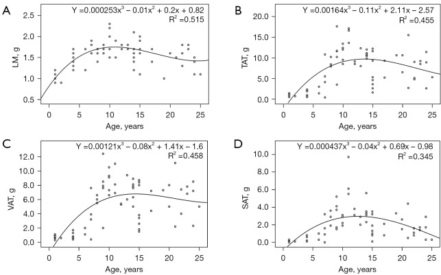
A total of 72 healthy female cynomolgus monkeys, aged 1-25 years, were included in this study. The average lumbar vBMD of female cynomolgus monkeys increased with age until the age of 10 years, around which it reached peak bone mass, with a relatively marked decline after the age of 13 years. The ABLRs of female cynomolgus monkeys in the premenopausal (13-19 years) and postmenopausal age groups (20-25 years) were -4.9% and -21.2%, respectively. Ridge regression analysis showed that age and subcutaneous adipose tissue (SAT) contributed positively to the average lumbar vBMD in animals aged ≤10 years, whereas in animals aged >10 years, age contributed negatively to lumbar vBMD. The RMS-CV% (RMS-SD) of the lumbar vBMD measured using QCT ranged from 0.47% to 1.60% (1.91-6.31 mg/cm).

CONCLUSIONS

Age-related changes in lumbar vBMD measured using QCT in healthy female monkeys showed similar trends to those in humans. Age and SAT may affect the lumbar vBMD in female cynomolgus monkeys. QCT revealed good precision in measuring the lumbar vBMD in female cynomolgus monkeys.

摘要

背景

食蟹猴广泛应用于骨质疏松相关研究,目前尚无证据表明使用定量计算机断层扫描(QCT)测量非人灵长类动物的体积骨密度(vBMD)存在与年龄相关的变化。本研究旨在描述腰椎vBMD特征随年龄的变化,分析腰椎vBMD与身体成分之间的关系,并研究QCT测量健康雌性食蟹猴的精度。

方法

使用三次回归模型描述腰椎vBMD的年龄相关变化,并计算腰椎的累积骨丢失率(ABLR)。采用Spearman等级相关和岭回归分析来研究平均腰椎vBMD与身体成分的关系。选择30只动物分析QCT测量的短期精度。精度用均方根变异系数(RMS-CV%)或均方根标准差(RMS-SD)表示。

结果

本研究共纳入72只年龄为1至25岁的健康雌性食蟹猴。雌性食蟹猴的平均腰椎vBMD随年龄增长而增加,直至10岁左右达到峰值骨量,13岁后相对明显下降。绝经前(13至19岁)和绝经后年龄组(20至25岁)雌性食蟹猴的ABLR分别为-4.9%和-21.2%。岭回归分析表明,年龄和皮下脂肪组织(SAT)对年龄≤10岁动物的平均腰椎vBMD有正向贡献,而在年龄>10岁的动物中,年龄对腰椎vBMD有负向贡献。使用QCT测量腰椎vBMD的RMS-CV%(RMS-SD)范围为0.47%至1.60%(1.91至6.31mg/cm)。

结论

使用QCT测量健康雌性食蟹猴腰椎vBMD的年龄相关变化与人类相似。年龄和SAT可能影响雌性食蟹猴的腰椎vBMD。QCT在测量雌性食蟹猴腰椎vBMD方面显示出良好的精度。

https://cdn.ncbi.nlm.nih.gov/pmc/blobs/4576/10102742/7982047dd80b/qims-13-04-2038-f2.jpg
https://cdn.ncbi.nlm.nih.gov/pmc/blobs/4576/10102742/6c9def9dbbb1/qims-13-04-2038-f1.jpg
https://cdn.ncbi.nlm.nih.gov/pmc/blobs/4576/10102742/7982047dd80b/qims-13-04-2038-f2.jpg
https://cdn.ncbi.nlm.nih.gov/pmc/blobs/4576/10102742/6c9def9dbbb1/qims-13-04-2038-f1.jpg
https://cdn.ncbi.nlm.nih.gov/pmc/blobs/4576/10102742/7982047dd80b/qims-13-04-2038-f2.jpg

相似文献

1
Age-related changes in lumbar bone mineral density measured using quantitative computed tomography in healthy female cynomolgus monkeys.使用定量计算机断层扫描测量健康雌性食蟹猴腰椎骨密度的年龄相关变化。
Quant Imaging Med Surg. 2023 Apr 1;13(4):2038-2052. doi: 10.21037/qims-22-763. Epub 2023 Feb 7.
2
Correlation analysis of positron emission tomography/computed tomography-magnetic resonance imaging of cannabinoid type 1 receptor in the lumbar spine and brain of aged osteoporosis female cynomolgus monkeys.老年骨质疏松雌性食蟹猴腰椎和脑内大麻素1型受体的正电子发射断层扫描/计算机断层扫描-磁共振成像相关性分析
Quant Imaging Med Surg. 2023 Dec 1;13(12):7924-7935. doi: 10.21037/qims-23-118. Epub 2023 Nov 9.
3
The precision study of dual energy X-ray absorptiometry for bone mineral density and body composition measurements in female cynomolgus monkeys.双能X线吸收法用于雌性食蟹猴骨密度和身体成分测量的精密度研究。
Quant Imaging Med Surg. 2022 Mar;12(3):2051-2057. doi: 10.21037/qims-21-799.
4
Lumbar Bone Mineral Density Phantomless Computed Tomography Measurements and Correlation with Age and Fracture Incidence.腰椎骨密度无体模计算机断层扫描测量及其与年龄和骨折发生率的相关性
Traffic Inj Prev. 2015;16 Suppl 2(0 2):S153-60. doi: 10.1080/15389588.2015.1054029.
5
Postoperative decrease of regional volumetric bone mineral density measured by quantitative computed tomography after lumbar fusion surgery in adjacent vertebrae.腰椎融合术后相邻节段定量 CT 测量的区域性容积骨密度术后下降。
Osteoporos Int. 2020 Jun;31(6):1163-1171. doi: 10.1007/s00198-020-05367-3. Epub 2020 Mar 13.
6
The impact of degenerative disc disease on regional volumetric bone mineral density (vBMD) measured by quantitative computed tomography.定量计算机断层扫描测量退行性椎间盘疾病对区域性容积骨密度(vBMD)的影响。
Spine J. 2020 Feb;20(2):181-190. doi: 10.1016/j.spinee.2019.02.017. Epub 2019 May 21.
7
Automated Opportunistic Osteoporosis Screening in Routine Computed Tomography of the Spine: Comparison With Dedicated Quantitative CT.脊柱常规计算机断层扫描中的自动机会性骨质疏松筛查:与专用定量CT的比较。
J Bone Miner Res. 2022 Jul;37(7):1287-1296. doi: 10.1002/jbmr.4575. Epub 2022 Jun 15.
8
Relationship of adiposity to bone volumetric density and microstructure in men and women across the adult lifespan.不论男女,在整个成年期,肥胖与骨体积密度和微观结构的关系。
Bone. 2013 Jul;55(1):119-25. doi: 10.1016/j.bone.2013.02.006. Epub 2013 Feb 18.
9
Quantitative computed tomography discriminates between postmenopausal women with low spine bone mineral density with vertebral fractures and those with low spine bone mineral density only: the SHATTER study.定量计算机断层扫描可区分伴有椎体骨折的绝经后低骨密度女性和仅有低骨密度的绝经后女性:SHATTER 研究。
Osteoporos Int. 2020 Apr;31(4):667-675. doi: 10.1007/s00198-020-05317-z. Epub 2020 Jan 28.
10
Correction of QCT vBMD using MRI measurements of marrow adipose tissue.利用骨髓脂肪组织 MRI 测量值校正 QCT vBMD。
Bone. 2019 Mar;120:504-511. doi: 10.1016/j.bone.2018.12.015. Epub 2018 Dec 21.

引用本文的文献

1
Correlation analysis of positron emission tomography/computed tomography-magnetic resonance imaging of cannabinoid type 1 receptor in the lumbar spine and brain of aged osteoporosis female cynomolgus monkeys.老年骨质疏松雌性食蟹猴腰椎和脑内大麻素1型受体的正电子发射断层扫描/计算机断层扫描-磁共振成像相关性分析
Quant Imaging Med Surg. 2023 Dec 1;13(12):7924-7935. doi: 10.21037/qims-23-118. Epub 2023 Nov 9.

本文引用的文献

1
Global, regional prevalence, and risk factors of osteoporosis according to the World Health Organization diagnostic criteria: a systematic review and meta-analysis.根据世界卫生组织诊断标准的全球、区域骨质疏松症患病率及危险因素:一项系统评价和荟萃分析
Osteoporos Int. 2022 Oct;33(10):2137-2153. doi: 10.1007/s00198-022-06454-3. Epub 2022 Jun 10.
2
Age, Sex, and Race/Ethnicity Associations between Fat Mass and Lean Mass with Bone Mineral Density: NHANES Data.年龄、性别和种族/民族与骨密度的脂肪量和瘦体量的关系:NHANES 数据。
Int J Environ Res Public Health. 2021 Nov 30;18(23):12606. doi: 10.3390/ijerph182312606.
3
Application of bone turnover markers and DXA and QCT in an elderly Chinese male population.
骨转换标志物及 DXA 和 QCT 在老年中国男性人群中的应用。
Ann Palliat Med. 2021 Jun;10(6):6351-6358. doi: 10.21037/apm-21-612.
4
Opportunistic Screening Using Low-Dose CT and the Prevalence of Osteoporosis in China: A Nationwide, Multicenter Study.利用低剂量 CT 进行机会性筛查与中国骨质疏松症的患病率:一项全国性、多中心研究。
J Bone Miner Res. 2021 Mar;36(3):427-435. doi: 10.1002/jbmr.4187. Epub 2020 Nov 4.
5
Lumbar bone mineral density measured by quantitative computed tomography (QCT): association with abdominal adipose tissue in different menopausal periods of Chinese women.采用定量计算机断层扫描(QCT)测量腰椎骨密度:与中国不同绝经阶段女性腹部脂肪组织的关系。
Gynecol Endocrinol. 2021 Mar;37(3):264-268. doi: 10.1080/09513590.2020.1836147. Epub 2020 Oct 21.
6
Chinese expert consensus on the diagnosis of osteoporosis by imaging and bone mineral density.中国骨质疏松症影像学与骨密度诊断专家共识
Quant Imaging Med Surg. 2020 Oct;10(10):2066-2077. doi: 10.21037/qims-2020-16.
7
Age-related fatty infiltration of lumbar paraspinal muscles: a normative reference database study in 516 Chinese females.腰椎旁肌肉的年龄相关性脂肪浸润:一项对516名中国女性的规范性参考数据库研究
Quant Imaging Med Surg. 2020 Aug;10(8):1590-1601. doi: 10.21037/qims-19-835.
8
Body compositions differently contribute to BMD in different age and gender: a pilot study by QCT.体成分在不同年龄和性别中对 BMD 的贡献不同:一项 QCT 研究的初步结果。
Arch Osteoporos. 2019 Mar 7;14(1):31. doi: 10.1007/s11657-019-0574-5.
9
Weekly teriparatide treatment increases vertebral body strength by improving cortical shell architecture in ovariectomized cynomolgus monkeys.每周特立帕肽治疗可通过改善去卵巢食蟹猴的皮质壳结构来增加椎体强度。
Bone. 2019 Apr;121:80-88. doi: 10.1016/j.bone.2019.01.006. Epub 2019 Jan 8.
10
Cortical bone mineral density is increased by the cathepsin K inhibitor ONO-5334, which leads to a robust increase in bone strength: results from a 16-month study in ovariectomised cynomolgus monkeys.破骨细胞 Cathepsin K 抑制剂 ONO-5334 可增加皮质骨骨密度,从而显著增强骨强度:在去卵巢食蟹猴中开展的为期 16 个月的研究结果。
J Bone Miner Metab. 2019 Jul;37(4):636-647. doi: 10.1007/s00774-018-0968-2. Epub 2018 Oct 24.