Helmholtz-Zentrum Dresden-Rossendorf, PET Center, Institute of Radiopharmaceutical Cancer Research, Bautzner Landstrasse 400, 01328, Dresden, Germany.
Department of Radiation Oncology, Charité - Universitätsmedizin Berlin, corporate member of Freie Universität Berlin and Humboldt-Universität zu Berlin, Berlin, Germany.
Eur J Nucl Med Mol Imaging. 2023 Jul;50(9):2751-2766. doi: 10.1007/s00259-023-06197-1. Epub 2023 Apr 20.
PET-derived metabolic tumor volume (MTV) and total lesion glycolysis of the primary tumor are known to be prognostic of clinical outcome in head and neck cancer (HNC). Including evaluation of lymph node metastases can further increase the prognostic value of PET but accurate manual delineation and classification of all lesions is time-consuming and prone to interobserver variability. Our goal, therefore, was development and evaluation of an automated tool for MTV delineation/classification of primary tumor and lymph node metastases in PET/CT investigations of HNC patients.
Automated lesion delineation was performed with a residual 3D U-Net convolutional neural network (CNN) incorporating a multi-head self-attention block. 698 [Formula: see text]F]FDG PET/CT scans from 3 different sites and 5 public databases were used for network training and testing. An external dataset of 181 [Formula: see text]F]FDG PET/CT scans from 2 additional sites was employed to assess the generalizability of the network. In these data, primary tumor and lymph node (LN) metastases were interactively delineated and labeled by two experienced physicians. Performance of the trained network models was assessed by 5-fold cross-validation in the main dataset and by pooling results from the 5 developed models in the external dataset. The Dice similarity coefficient (DSC) for individual delineation tasks and the primary tumor/metastasis classification accuracy were used as evaluation metrics. Additionally, a survival analysis using univariate Cox regression was performed comparing achieved group separation for manual and automated delineation, respectively.
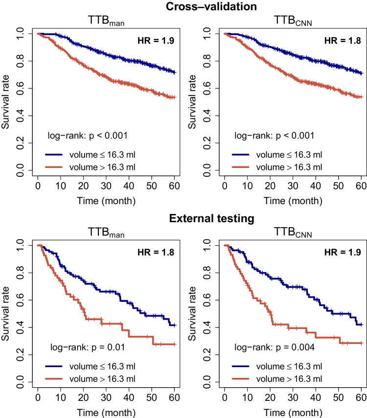
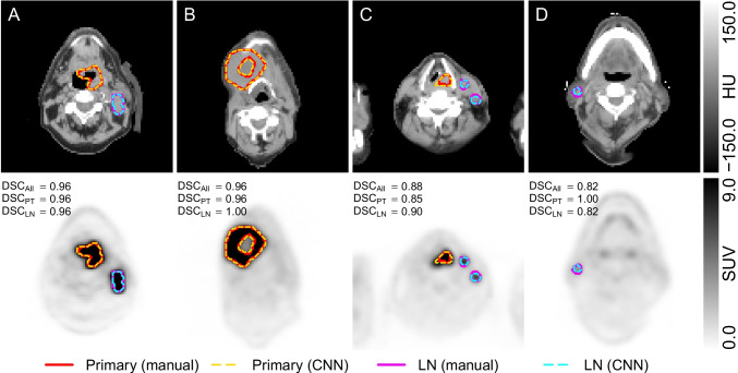
In the cross-validation experiment, delineation of all malignant lesions with the trained U-Net models achieves DSC of 0.885, 0.805, and 0.870 for primary tumor, LN metastases, and the union of both, respectively. In external testing, the DSC reaches 0.850, 0.724, and 0.823 for primary tumor, LN metastases, and the union of both, respectively. The voxel classification accuracy was 98.0% and 97.9% in cross-validation and external data, respectively. Univariate Cox analysis in the cross-validation and the external testing reveals that manually and automatically derived total MTVs are both highly prognostic with respect to overall survival, yielding essentially identical hazard ratios (HR) ([Formula: see text]; [Formula: see text] vs. [Formula: see text]; [Formula: see text] in cross-validation and [Formula: see text]; [Formula: see text] vs. [Formula: see text]; [Formula: see text] in external testing).
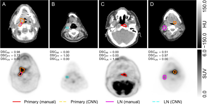
To the best of our knowledge, this work presents the first CNN model for successful MTV delineation and lesion classification in HNC. In the vast majority of patients, the network performs satisfactory delineation and classification of primary tumor and lymph node metastases and only rarely requires more than minimal manual correction. It is thus able to massively facilitate study data evaluation in large patient groups and also does have clear potential for supervised clinical application.
正电子发射断层扫描(PET)衍生的代谢肿瘤体积(MTV)和原发肿瘤总肿瘤糖酵解已被证明对头颈癌(HNC)的临床结果具有预后价值。包括对淋巴结转移的评估可以进一步提高 PET 的预后价值,但对所有病变的准确手动描绘和分类既耗时又容易出现观察者间的变异性。因此,我们的目标是开发和评估一种用于 HNC 患者 PET/CT 研究中原发性肿瘤和淋巴结转移的 MTV 描绘/分类的自动化工具。
使用包含多头自注意块的剩余 3D U-Net 卷积神经网络(CNN)进行自动病变描绘。来自 3 个不同地点和 5 个公共数据库的 698 个 [Formula: see text]FDG PET/CT 扫描用于网络训练和测试。另外 2 个地点的 181 个 [Formula: see text]FDG PET/CT 扫描的外部数据集用于评估网络的泛化能力。在这些数据中,由两位经验丰富的医生对原发肿瘤和淋巴结(LN)转移进行交互式描绘和标记。在主要数据集的 5 折交叉验证中评估训练后的网络模型的性能,并在外部数据集的 5 个开发模型中汇总结果。使用 Dice 相似系数(DSC)作为评估指标,分别评估单个描绘任务的 DSC 和原发肿瘤/转移分类准确性。此外,还使用单变量 Cox 回归进行了生存分析,比较了手动和自动描绘分别实现的组分离。
在交叉验证实验中,使用训练的 U-Net 模型对所有恶性病变进行描绘,分别达到原发性肿瘤、LN 转移和两者联合的 DSC 为 0.885、0.805 和 0.870。在外部测试中,分别达到原发性肿瘤、LN 转移和两者联合的 DSC 为 0.850、0.724 和 0.823。在交叉验证和外部数据中,体素分类准确率分别为 98.0%和 97.9%。交叉验证和外部测试中的单变量 Cox 分析表明,手动和自动推导的总 MTV 均与总生存率高度相关,产生的危险比(HR)基本相同([Formula: see text];[Formula: see text]与 [Formula: see text];[Formula: see text]在交叉验证和 [Formula: see text];[Formula: see text]与 [Formula: see text];[Formula: see text]在外部测试中)。
据我们所知,这项工作首次提出了用于成功描绘 HNC 中 MTV 和病变分类的 CNN 模型。在绝大多数患者中,网络对原发性肿瘤和淋巴结转移的描绘和分类表现令人满意,仅在极少数情况下需要进行超过最小限度的手动修正。因此,它能够极大地促进大型患者组的研究数据评估,并且对于有监督的临床应用也具有明显的潜力。