Radiation Biology Research Center, Iran University of Medical Science (IUMS), Tehran, Iran.
Radiation Science Department, Iran University of Medical Science (IUMS), Tehran, Iran.
IET Nanobiotechnol. 2023 May;17(3):212-223. doi: 10.1049/nbt2.12129. Epub 2023 Apr 21.
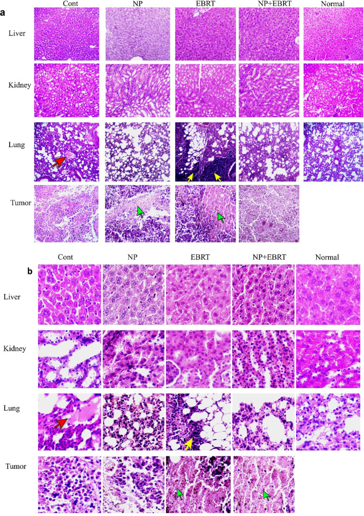
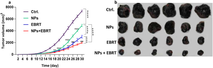
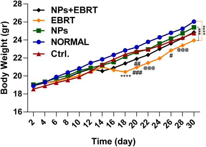
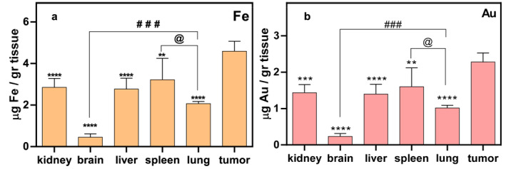
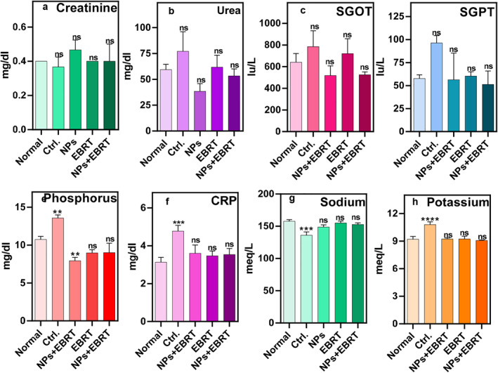
Melanoma is a dangerous type of skin cancer sometimes treated with radiotherapy. However, it induces damage to the surrounding healthy tissue and possibly further away areas. Therefore, it is necessary to give a lower dose to the patient with targeted therapy. In this study, the radio-sensitising effect of gold-coated iron oxide nanoparticles on electron beam radiotherapy of a melanoma tumour with magnetic targeting in a mouse model was investigated. Gold-coated iron oxide nanoparticles were prepared in a steady procedure. The melanoma tumour model was induced in mice. Animals were divided into five groups: (1) normal; (2) melanoma; (3) gold-coated iron oxide nanoparticles alone; (4) electron beam radiotherapy; (5) electron beam radiotherapy plus gold-coated iron oxide nanoparticles. The magnet was placed on the tumour site for 2 h. The tumours were then exposed to 6 MeV electron beam radiotherapy for a dose of 8 Gy. Inductively coupled plasma optical emission spectrometry test, hematoxylin and eosin staining, and enzyme-linked immunosorbent assay blood test were also performed. Gold-coated iron oxide nanoparticles with magnetic targeting before electron beam radiotherapy reduced the growth of the tumour compared to the control group. Blood tests did not show any significant toxicity. Deposition of nanoparticles was more in the tumour and spleen tissue and to a lesser extent in the liver, kidney, and lung tissues. The synergistic effect of nanoparticles administered by the intraperitoneal route and then concentrated into the tumour area by application of an external permanent magnet, before delivery of the electron beam radiotherapy improved the overall cancer treatment outcome and prevented metal distribution side effects.
黑色素瘤是一种危险的皮肤癌,有时采用放射疗法治疗。然而,放射疗法会对周围健康组织造成损伤,甚至可能对更远的区域造成损伤。因此,有必要通过靶向治疗为患者给予更低的剂量。在这项研究中,研究了金包覆氧化铁纳米粒子对电子束放射疗法治疗小鼠模型中黑色素瘤肿瘤的放射增敏作用,并进行了磁性靶向。金包覆氧化铁纳米粒子是通过稳定的程序制备的。在小鼠中诱导黑色素瘤肿瘤模型。动物分为五组:(1)正常;(2)黑色素瘤;(3)金包覆氧化铁纳米粒子单独组;(4)电子束放射疗法组;(5)电子束放射疗法加金包覆氧化铁纳米粒子组。将磁铁放置在肿瘤部位 2 小时。然后,将肿瘤暴露于 6 MeV 电子束放射疗法下,剂量为 8 Gy。还进行了电感耦合等离子体发射光谱测试、苏木精和伊红染色以及酶联免疫吸附测定血液测试。与对照组相比,电子束放射疗法前进行磁性靶向的金包覆氧化铁纳米粒子可减少肿瘤的生长。血液测试未显示出任何明显的毒性。纳米粒子的沉积更多地发生在肿瘤和脾脏组织中,而在肝脏、肾脏和肺部组织中则较少。通过腹腔途径给予纳米粒子并在应用外部永磁体将其集中到肿瘤区域后,与电子束放射疗法联合使用,可提高整体癌症治疗效果并防止金属分布的副作用。