Institute of Tissue Medicine and Pathology, University of Bern, Murtenstrasse 31, CH-3008, Bern, Switzerland.
Department of Otorhinolaryngology, Head and Neck Surgery, Inselspital, Bern University Hospital, and University of Bern, Bern, Switzerland.
Head Neck Pathol. 2023 Sep;17(3):803-807. doi: 10.1007/s12105-023-01554-w. Epub 2023 Apr 28.
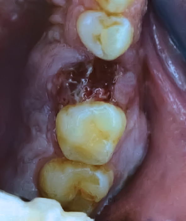
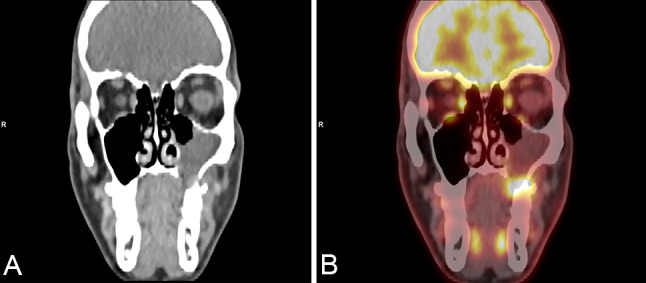
In recent years, the list of tumor entities in the sinonasal tract has significantly expanded, requiring advanced diagnostic testing. We report the case of a 32-year-old patient with an unusual NUT carcinoma originating in the maxillary sinus, which showed extensive well-differentiated, papillary squamous morphology, similar to the spectrum of the recently described DEK::AFF2 fusion-associated carcinoma.
We performed immunohistochemical and molecular studies including EBV- and HPV-testing, as well as DNA/RNA next generation sequencing.
The tumor showed predominantly exophytic papillary growth with mature squamous differentiation. An additional component harbored atypical, less differentiated basaloid tumor cells with infiltration of the adjacent stroma. Conspicuous inflammation was evident. There was no evidence of HPV DNA or EBV RNA. Next-generation sequencing revealed a NUT::NSD3 gene fusion corresponding to ("speckled-type") immunopositivity of NUT in the tumor cells.
We describe a NUT::NSD3 gene fusion-associated NUT carcinoma of the sinonasal tract with a deceptively well-differentiated papillary growth pattern, thus expanding the morphological spectrum of this typically poorly differentiated neoplasm.
近年来,鼻窦道肿瘤实体的清单显著扩大,需要进行高级诊断测试。我们报告了一例 32 岁患者的罕见 NUT 癌,起源于上颌窦,其表现出广泛的良好分化、乳头状鳞状形态,类似于最近描述的 DEK::AFF2 融合相关癌的范围。
我们进行了免疫组织化学和分子研究,包括 EBV-和 HPV 检测,以及 DNA/RNA 下一代测序。
肿瘤主要表现为外生性乳头状生长,具有成熟的鳞状分化。另一个成分含有不典型的、分化程度较低的基底样肿瘤细胞,伴有相邻基质的浸润。明显的炎症是明显的。没有 HPV DNA 或 EBV RNA 的证据。下一代测序显示 NUT::NSD3 基因融合,对应于肿瘤细胞中 NUT 的(“斑点型”)免疫阳性。
我们描述了一种鼻窦道的 NUT::NSD3 基因融合相关的 NUT 癌,具有欺骗性的良好分化的乳头状生长模式,从而扩展了这种典型的低分化肿瘤的形态谱。