Department of Anatomy, Korea University College of Medicine, Seoul, 02841, Republic of Korea.
Department of Bio-Information Science, Ewha Womans University, Seoul, 03760, Republic of Korea.
Adv Sci (Weinh). 2023 Jul;10(20):e2301787. doi: 10.1002/advs.202301787. Epub 2023 May 12.
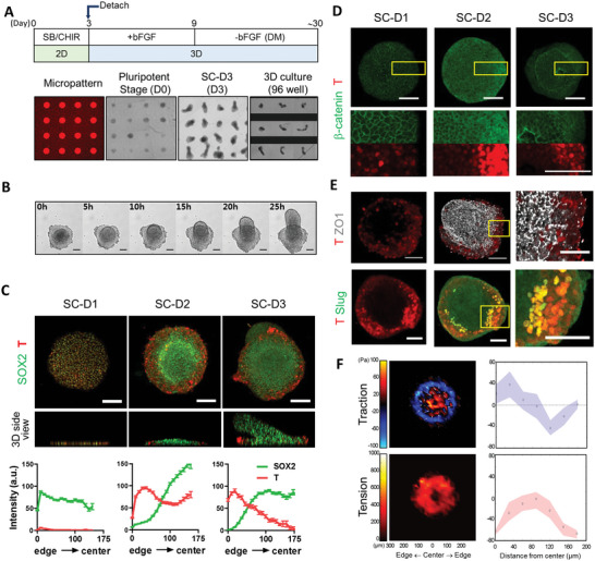
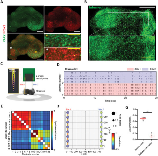
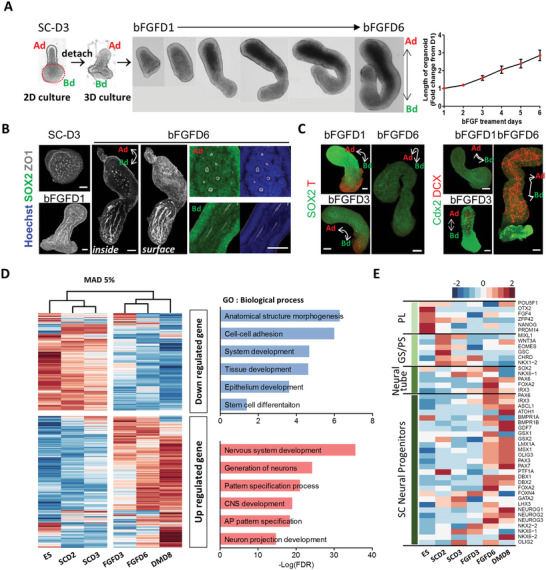
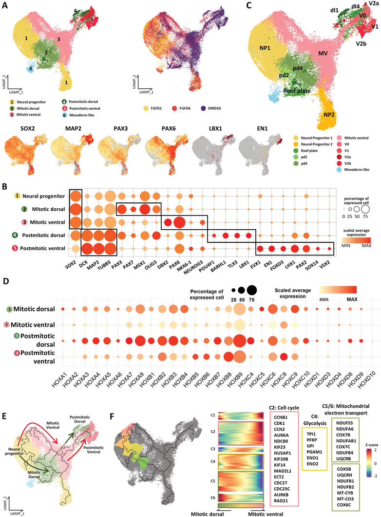
Axis formation and related spatial patterning are initiated by symmetry breaking during development. A geometrically confined culture of human pluripotent stem cells (hPSCs) mimics symmetry breaking and cell patterning. Using this, polarized spinal cord organoids (pSCOs) with a self-organized dorsoventral (DV) organization are generated. The application of caudalization signals promoted regionalized cell differentiation along the radial axis and protrusion morphogenesis in confined hPSC colonies. These detached colonies grew into extended spinal cord-like organoids, which established self-ordered DV patterning along the long axis through the spontaneous expression of polarized DV patterning morphogens. The proportions of dorsal/ventral domains in the pSCOs can be controlled by the changes in the initial size of micropatterns, which altered the ratio of center-edge cells in 2D. In mature pSCOs, highly synchronized neural activity is separately detected in the dorsal and ventral side, indicating functional as well as structural patterning established in the organoids. This study provides a simple and precisely controllable method to generate spatially ordered organoids for the understanding of the biological principles of cell patterning and axis formation during neural development.
轴形成和相关的空间模式是通过发育过程中的对称性破缺启动的。人类多能干细胞(hPSC)的几何限制培养模拟了对称性破缺和细胞模式形成。利用这种方法,可以生成具有自组织背腹(DV)组织的极化脊髓类器官(pSCO)。尾侧化信号的应用促进了沿着辐射轴的区域化细胞分化,并在受限的 hPSC 集落中促进了突起形态发生。这些分离的集落生长成延伸的脊髓样类器官,通过极化的 DV 模式形成形态发生因子的自发表达,沿着长轴建立了自组织的 DV 模式。通过改变初始微图案的大小,可以控制 pSCO 中背/腹区域的比例,从而改变 2D 中的中心-边缘细胞的比例。在成熟的 pSCO 中,在背部和腹部可以分别检测到高度同步的神经活动,表明在类器官中建立了功能和结构模式。这项研究提供了一种简单且可精确控制的方法来生成空间有序的类器官,以了解神经发育过程中细胞模式形成和轴形成的生物学原理。