• 文献检索
  • 文档翻译
  • 深度研究
  • 学术资讯
  • Suppr Zotero 插件Zotero 插件
  • 邀请有礼
  • 套餐&价格
  • 历史记录
应用&插件
Suppr Zotero 插件Zotero 插件浏览器插件Mac 客户端Windows 客户端微信小程序
定价
高级版会员购买积分包购买API积分包
服务
文献检索文档翻译深度研究API 文档MCP 服务
关于我们
关于 Suppr公司介绍联系我们用户协议隐私条款
关注我们

Suppr 超能文献

核心技术专利:CN118964589B侵权必究
粤ICP备2023148730 号-1Suppr @ 2026

文献检索

告别复杂PubMed语法,用中文像聊天一样搜索,搜遍4000万医学文献。AI智能推荐,让科研检索更轻松。

立即免费搜索

文件翻译

保留排版,准确专业,支持PDF/Word/PPT等文件格式,支持 12+语言互译。

免费翻译文档

深度研究

AI帮你快速写综述,25分钟生成高质量综述,智能提取关键信息,辅助科研写作。

立即免费体验

针对局部区域性乳腺癌放疗的深度学习分割模型在两个不同数据集上训练后的输出比较。

Comparison of the output of a deep learning segmentation model for locoregional breast cancer radiotherapy trained on 2 different datasets.

作者信息

Bakx Nienke, van der Sangen Maurice, Theuws Jacqueline, Bluemink Hanneke, Hurkmans Coen

机构信息

Catharina Hospital, Department of Radiation Oncology, 5602ZA Eindhoven, the Netherlands.

Technical University Eindhoven, Faculties of Physics and Electrical Engineering, 5600MB Eindhoven, the Netherlands.

出版信息

Tech Innov Patient Support Radiat Oncol. 2023 May 13;26:100209. doi: 10.1016/j.tipsro.2023.100209. eCollection 2023 Jun.

DOI:10.1016/j.tipsro.2023.100209
PMID:37213441
原文链接:https://pmc.ncbi.nlm.nih.gov/articles/PMC10199413/
Abstract

INTRODUCTION

The development of deep learning (DL) models for auto-segmentation is increasing and more models become commercially available. Mostly, commercial models are trained on external data. To study the effect of using a model trained on external data, compared to the same model trained on in-house collected data, the performance of these two DL models was evaluated.

METHODS

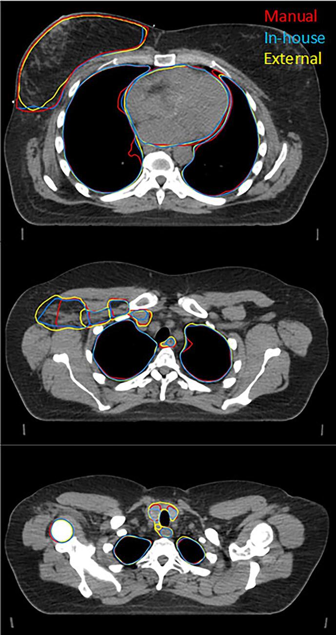
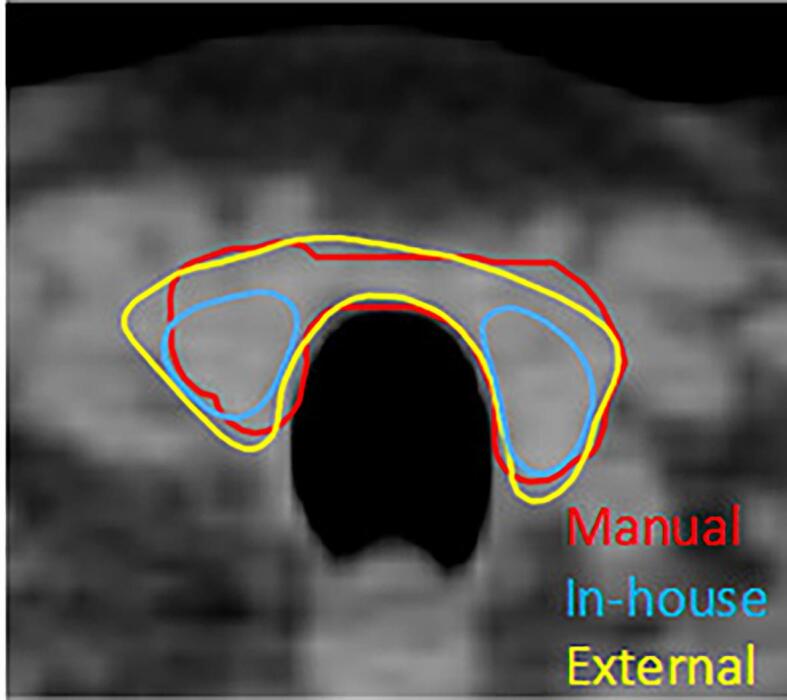
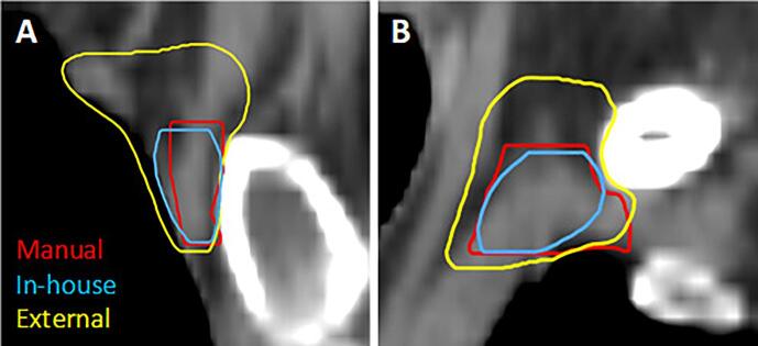
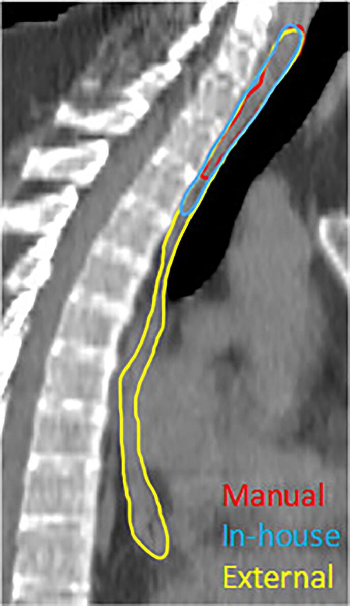
The evaluation was performed using in-house collected data of 30 breast cancer patients. Quantitative analysis was performed using Dice similarity coefficient (DSC), surface DSC (sDSC) and 95th percentile of Hausdorff Distance (95% HD). These values were compared with previously reported inter-observer variations (IOV).

RESULTS

For a number of structures, statistically significant differences were found between the two models. For organs at risk, mean values for DSC ranged from 0.63 to 0.98 and 0.71 to 0.96 for the in-house and external model, respectively. For target volumes, mean DSC values of 0.57 to 0.94 and 0.33 to 0.92 were found. The difference of 95% HD values ranged 0.08 to 3.23 mm between the two models, except for CTVn4 with 9.95 mm. For the external model, both DSC and 95% HD are outside the range of IOV for CTVn4, whereas this is the case for the DSC found for the thyroid of the in-house model.

CONCLUSIONS

Statistically significant differences were found between both models, which were mostly within published inter-observer variations, showing clinical usefulness of both models. Our findings could encourage discussion and revision of existing guidelines, to further decrease inter-observer, but also inter-institute variability.

摘要

引言

用于自动分割的深度学习(DL)模型不断发展,越来越多的模型开始商业化。大多数情况下,商业模型是基于外部数据进行训练的。为了研究使用基于外部数据训练的模型与基于内部收集数据训练的相同模型相比的效果,对这两种DL模型的性能进行了评估。

方法

使用内部收集的30例乳腺癌患者的数据进行评估。采用骰子相似系数(DSC)、表面DSC(sDSC)和95% 豪斯多夫距离(95% HD)进行定量分析。将这些值与先前报道的观察者间差异(IOV)进行比较。

结果

在多个结构上,发现两种模型之间存在统计学显著差异。对于危及器官,内部模型和外部模型的DSC平均值分别为0.63至0.98和0.71至0.96。对于靶区体积,发现平均DSC值分别为0.57至0.94和0.33至0.92。除CTVn4为9.95mm外,两种模型的95% HD值差异范围为0.08至3.23mm。对于外部模型,CTVn4的DSC和95% HD均超出IOV范围,而内部模型甲状腺的DSC情况也是如此。

结论

两种模型之间存在统计学显著差异,大多在已发表的观察者间差异范围内,表明两种模型均具有临床实用性。我们的研究结果可能会促使对现有指南进行讨论和修订,以进一步减少观察者间以及机构间的变异性。

https://cdn.ncbi.nlm.nih.gov/pmc/blobs/ccd6/10199413/44fea31b6901/gr4.jpg
https://cdn.ncbi.nlm.nih.gov/pmc/blobs/ccd6/10199413/ac36151d1185/gr1.jpg
https://cdn.ncbi.nlm.nih.gov/pmc/blobs/ccd6/10199413/67b6c928f08b/gr2.jpg
https://cdn.ncbi.nlm.nih.gov/pmc/blobs/ccd6/10199413/a56983492be1/gr3.jpg
https://cdn.ncbi.nlm.nih.gov/pmc/blobs/ccd6/10199413/44fea31b6901/gr4.jpg
https://cdn.ncbi.nlm.nih.gov/pmc/blobs/ccd6/10199413/ac36151d1185/gr1.jpg
https://cdn.ncbi.nlm.nih.gov/pmc/blobs/ccd6/10199413/67b6c928f08b/gr2.jpg
https://cdn.ncbi.nlm.nih.gov/pmc/blobs/ccd6/10199413/a56983492be1/gr3.jpg
https://cdn.ncbi.nlm.nih.gov/pmc/blobs/ccd6/10199413/44fea31b6901/gr4.jpg

相似文献

1
Comparison of the output of a deep learning segmentation model for locoregional breast cancer radiotherapy trained on 2 different datasets.针对局部区域性乳腺癌放疗的深度学习分割模型在两个不同数据集上训练后的输出比较。
Tech Innov Patient Support Radiat Oncol. 2023 May 13;26:100209. doi: 10.1016/j.tipsro.2023.100209. eCollection 2023 Jun.
2
Comparison of the use of a clinically implemented deep learning segmentation model with the simulated study setting for breast cancer patients receiving radiotherapy.比较在接受放射治疗的乳腺癌患者中使用临床实施的深度学习分割模型与模拟研究环境的效果。
Acta Oncol. 2024 Jun 20;63:477-481. doi: 10.2340/1651-226X.2024.34986.
3
Transfer learning for auto-segmentation of 17 organs-at-risk in the head and neck: Bridging the gap between institutional and public datasets.基于迁移学习的头颈部 17 个危及器官自动分割:弥合机构数据集和公共数据集之间的差距。
Med Phys. 2024 Jul;51(7):4767-4777. doi: 10.1002/mp.16997. Epub 2024 Feb 20.
4
Assessment of deep learning-based auto-contouring on interobserver consistency in target volume and organs-at-risk delineation for breast cancer: Implications for RTQA program in a multi-institutional study.基于深度学习的自动勾画在乳腺癌靶区和危及器官勾画中观察者间一致性的评估:多中心研究中对 RTQA 计划的影响。
Breast. 2024 Feb;73:103599. doi: 10.1016/j.breast.2023.103599. Epub 2023 Nov 15.
5
Training, validation, and clinical implementation of a deep-learning segmentation model for radiotherapy of loco-regional breast cancer.深度学习分割模型在局部区域性乳腺癌放射治疗中的训练、验证和临床应用。
Radiother Oncol. 2022 Aug;173:62-68. doi: 10.1016/j.radonc.2022.05.018. Epub 2022 May 23.
6
Clinical feasibility of deep learning-based auto-segmentation of target volumes and organs-at-risk in breast cancer patients after breast-conserving surgery.保乳手术后乳腺癌患者基于深度学习的靶区体积和危及器官自动分割的临床可行性
Radiat Oncol. 2021 Feb 25;16(1):44. doi: 10.1186/s13014-021-01771-z.
7
Comparing deep learning-based auto-segmentation of organs at risk and clinical target volumes to expert inter-observer variability in radiotherapy planning.比较基于深度学习的危及器官和临床靶区自动分割与放射治疗计划中专家间观察者变异性。
Radiother Oncol. 2020 Mar;144:152-158. doi: 10.1016/j.radonc.2019.10.019. Epub 2019 Dec 5.
8
Application of simultaneous uncertainty quantification for image segmentation with probabilistic deep learning: Performance benchmarking of oropharyngeal cancer target delineation as a use-case.概率深度学习在图像分割中的同步不确定性量化应用:以口咽癌靶区勾画为例的性能基准测试
medRxiv. 2023 Feb 24:2023.02.20.23286188. doi: 10.1101/2023.02.20.23286188.
9
Comparative clinical evaluation of atlas and deep-learning-based auto-segmentation of organ structures in liver cancer.基于 atlas 和深度学习的肝癌器官结构自动分割的临床对比评估。
Radiat Oncol. 2019 Nov 27;14(1):213. doi: 10.1186/s13014-019-1392-z.
10
Clinical evaluation of a deep learning segmentation model including manual adjustments afterwards for locally advanced breast cancer.深度学习分割模型在局部晚期乳腺癌中的临床评估,后续包括手动调整。
Tech Innov Patient Support Radiat Oncol. 2023 May 13;26:100211. doi: 10.1016/j.tipsro.2023.100211. eCollection 2023 Jun.

引用本文的文献

1
Validation of different automated segmentation models for target volume contouring in postoperative radiotherapy for breast cancer and regional nodal irradiation.乳腺癌术后放疗及区域淋巴结照射中不同自动分割模型用于靶区轮廓勾画的验证
Clin Transl Radiat Oncol. 2024 Sep 11;49:100855. doi: 10.1016/j.ctro.2024.100855. eCollection 2024 Nov.
2
Feasibility study of adaptive radiotherapy with Ethos for breast cancer.使用Ethos进行乳腺癌自适应放疗的可行性研究。
Front Oncol. 2023 Nov 13;13:1274082. doi: 10.3389/fonc.2023.1274082. eCollection 2023.

本文引用的文献

1
Clinical evaluation of a deep learning segmentation model including manual adjustments afterwards for locally advanced breast cancer.深度学习分割模型在局部晚期乳腺癌中的临床评估,后续包括手动调整。
Tech Innov Patient Support Radiat Oncol. 2023 May 13;26:100211. doi: 10.1016/j.tipsro.2023.100211. eCollection 2023 Jun.
2
Training, validation, and clinical implementation of a deep-learning segmentation model for radiotherapy of loco-regional breast cancer.深度学习分割模型在局部区域性乳腺癌放射治疗中的训练、验证和临床应用。
Radiother Oncol. 2022 Aug;173:62-68. doi: 10.1016/j.radonc.2022.05.018. Epub 2022 May 23.
3
Machine Learning for Auto-Segmentation in Radiotherapy Planning.
机器学习在放射治疗计划中的自动分割。
Clin Oncol (R Coll Radiol). 2022 Feb;34(2):74-88. doi: 10.1016/j.clon.2021.12.003. Epub 2022 Jan 5.
4
Deep learning for segmentation in radiation therapy planning: a review.深度学习在放射治疗计划中的分割应用:综述
J Med Imaging Radiat Oncol. 2021 Aug;65(5):578-595. doi: 10.1111/1754-9485.13286. Epub 2021 Jul 26.
5
Clinically Applicable Segmentation of Head and Neck Anatomy for Radiotherapy: Deep Learning Algorithm Development and Validation Study.临床可应用的头颈部解剖结构勾画:放射治疗深度学习算法的开发与验证研究。
J Med Internet Res. 2021 Jul 12;23(7):e26151. doi: 10.2196/26151.
6
Clinical integration of machine learning for curative-intent radiation treatment of patients with prostate cancer.临床整合机器学习治疗前列腺癌患者的治疗意图放射治疗。
Nat Med. 2021 Jun;27(6):999-1005. doi: 10.1038/s41591-021-01359-w. Epub 2021 Jun 3.
7
Novel Autosegmentation Spatial Similarity Metrics Capture the Time Required to Correct Segmentations Better Than Traditional Metrics in a Thoracic Cavity Segmentation Workflow.新型自动分割空间相似性度量标准比传统度量标准更能准确反映分割所需时间,这在胸部腔室分割工作流程中表现得尤为明显。
J Digit Imaging. 2021 Jun;34(3):541-553. doi: 10.1007/s10278-021-00460-3. Epub 2021 May 23.
8
Geometric contour variation in clinical target volume of axillary lymph nodes in breast cancer radiotherapy: an AIRO multi-institutional study.乳腺癌放疗中腋窝淋巴结临床靶区的几何轮廓变化:AIRO 多机构研究。
Br J Radiol. 2021 Jul 1;94(1123):20201177. doi: 10.1259/bjr.20201177. Epub 2021 Apr 21.
9
Clinical feasibility of deep learning-based auto-segmentation of target volumes and organs-at-risk in breast cancer patients after breast-conserving surgery.保乳手术后乳腺癌患者基于深度学习的靶区体积和危及器官自动分割的临床可行性
Radiat Oncol. 2021 Feb 25;16(1):44. doi: 10.1186/s13014-021-01771-z.
10
Evaluation of measures for assessing time-saving of automatic organ-at-risk segmentation in radiotherapy.放射治疗中自动危及器官分割时间节省评估措施的评价
Phys Imaging Radiat Oncol. 2019 Dec 17;13:1-6. doi: 10.1016/j.phro.2019.12.001. eCollection 2020 Jan.