• 文献检索
  • 文档翻译
  • 深度研究
  • 学术资讯
  • Suppr Zotero 插件Zotero 插件
  • 邀请有礼
  • 套餐&价格
  • 历史记录
应用&插件
Suppr Zotero 插件Zotero 插件浏览器插件Mac 客户端Windows 客户端微信小程序
定价
高级版会员购买积分包购买API积分包
服务
文献检索文档翻译深度研究API 文档MCP 服务
关于我们
关于 Suppr公司介绍联系我们用户协议隐私条款
关注我们

Suppr 超能文献

核心技术专利:CN118964589B侵权必究
粤ICP备2023148730 号-1Suppr @ 2026

文献检索

告别复杂PubMed语法,用中文像聊天一样搜索,搜遍4000万医学文献。AI智能推荐,让科研检索更轻松。

立即免费搜索

文件翻译

保留排版,准确专业,支持PDF/Word/PPT等文件格式,支持 12+语言互译。

免费翻译文档

深度研究

AI帮你快速写综述,25分钟生成高质量综述,智能提取关键信息,辅助科研写作。

立即免费体验

严重急性呼吸综合征冠状病毒2型E蛋白的二聚体跨膜结构

Dimeric Transmembrane Structure of the SARS-CoV-2 E Protein.

作者信息

Zhang Rongfu, Qin Huajun, Prasad Ramesh, Fu Riqiang, Zhou Huan-Xiang, Cross Timothy A

机构信息

Department of Chemistry and Biochemistry, Florida State University, Tallahassee, FL 32306.

National High Magnetic Field Laboratory, Tallahassee, FL 32310.

出版信息

bioRxiv. 2023 May 8:2023.05.07.539752. doi: 10.1101/2023.05.07.539752.

DOI:10.1101/2023.05.07.539752
PMID:37214926
原文链接:https://pmc.ncbi.nlm.nih.gov/articles/PMC10197518/
Abstract

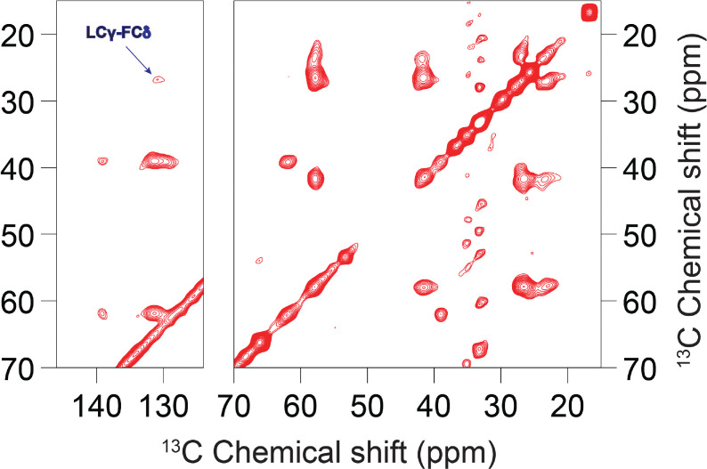
The SARS-CoV-2 E protein is a transmembrane (TM) protein with its N-terminus exposed on the external surface of the virus. Here, the TM structure of the E protein is characterized by oriented sample and magic angle spinning solid-state NMR in lipid bilayers and refined by molecular dynamics simulations. This protein has been found to be a pentamer, with a hydrophobic pore that appears to function as an ion channel. We identified only a symmetric helix-helix interface, leading to a dimeric structure that does not support channel activity. The two helices have a tilt angle of only 6°, resulting in an extended interface dominated by Leu and Val sidechains. While residues Val14-Thr35 are almost all buried in the hydrophobic region of the membrane, Asn15 lines a water-filled pocket that potentially serves as a drug-binding site. The E and other viral proteins may adopt different oligomeric states to help perform multiple functions.

摘要

严重急性呼吸综合征冠状病毒2(SARS-CoV-2)的E蛋白是一种跨膜(TM)蛋白,其N端暴露于病毒外表面。在此,通过在脂质双层中进行定向样品和魔角旋转固态核磁共振对E蛋白的跨膜结构进行了表征,并通过分子动力学模拟进行了优化。已发现该蛋白为五聚体,具有一个似乎起离子通道作用的疏水孔。我们仅鉴定出一个对称的螺旋-螺旋界面,导致形成不支持通道活性的二聚体结构。这两个螺旋的倾斜角仅为6°,形成了一个由亮氨酸和缬氨酸侧链主导的延伸界面。虽然缬氨酸14-苏氨酸35残基几乎全部埋在膜的疏水区域中,但天冬酰胺15排列在一个可能用作药物结合位点的充满水的口袋中。E蛋白和其他病毒蛋白可能采用不同的寡聚状态来帮助执行多种功能。

https://cdn.ncbi.nlm.nih.gov/pmc/blobs/1401/10197518/982aa4b47e16/nihpp-2023.05.07.539752v1-f0006.jpg
https://cdn.ncbi.nlm.nih.gov/pmc/blobs/1401/10197518/19d64763ea4f/nihpp-2023.05.07.539752v1-f0001.jpg
https://cdn.ncbi.nlm.nih.gov/pmc/blobs/1401/10197518/88adb3ef6e99/nihpp-2023.05.07.539752v1-f0002.jpg
https://cdn.ncbi.nlm.nih.gov/pmc/blobs/1401/10197518/7c381d791a39/nihpp-2023.05.07.539752v1-f0003.jpg
https://cdn.ncbi.nlm.nih.gov/pmc/blobs/1401/10197518/79e9bcfe90da/nihpp-2023.05.07.539752v1-f0004.jpg
https://cdn.ncbi.nlm.nih.gov/pmc/blobs/1401/10197518/d411e7166a3c/nihpp-2023.05.07.539752v1-f0005.jpg
https://cdn.ncbi.nlm.nih.gov/pmc/blobs/1401/10197518/982aa4b47e16/nihpp-2023.05.07.539752v1-f0006.jpg

相似文献

1
Dimeric Transmembrane Structure of the SARS-CoV-2 E Protein.严重急性呼吸综合征冠状病毒2型E蛋白的二聚体跨膜结构
bioRxiv. 2023 May 8:2023.05.07.539752. doi: 10.1101/2023.05.07.539752.
2
Dimeric Transmembrane Structure of the SARS-CoV-2 E Protein.SARS-CoV-2 刺突蛋白的二聚体跨膜结构。
Commun Biol. 2023 Nov 1;6(1):1109. doi: 10.1038/s42003-023-05490-x.
3
Structure of CrgA, a cell division structural and regulatory protein from Mycobacterium tuberculosis, in lipid bilayers.结核分枝杆菌细胞分裂结构与调节蛋白CrgA在脂质双分子层中的结构
Proc Natl Acad Sci U S A. 2015 Jan 13;112(2):E119-26. doi: 10.1073/pnas.1415908112. Epub 2014 Dec 29.
4
Structure and Drug Binding of the SARS-CoV-2 Envelope Protein in Phospholipid Bilayers.严重急性呼吸综合征冠状病毒2包膜蛋白在磷脂双分子层中的结构与药物结合
Res Sq. 2020 Sep 24:rs.3.rs-77124. doi: 10.21203/rs.3.rs-77124/v1.
5
Oligomeric State and Drug Binding of the SARS-CoV-2 Envelope Protein Are Sensitive to the Ectodomain.SARS-CoV-2 包膜蛋白的寡聚状态和药物结合对其胞外域敏感。
J Am Chem Soc. 2024 Sep 4;146(35):24537-24552. doi: 10.1021/jacs.4c07686. Epub 2024 Aug 21.
6
Impact of histidine residues on the transmembrane helices of viroporins.组氨酸残基对病毒离子通道跨膜螺旋的影响。
Mol Membr Biol. 2013 Nov;30(7):360-9. doi: 10.3109/09687688.2013.842657. Epub 2013 Oct 9.
7
Molecular dynamics simulations of the dimerization of transmembrane alpha-helices.跨膜α-螺旋二聚体的分子动力学模拟。
Acc Chem Res. 2010 Mar 16;43(3):388-96. doi: 10.1021/ar900211k.
8
Hexamethylene amiloride binds the SARS-CoV-2 envelope protein at the protein-lipid interface.六亚甲基甲脒与 SARS-CoV-2 包膜蛋白在蛋白-脂双层界面结合。
Protein Sci. 2023 Oct;32(10):e4755. doi: 10.1002/pro.4755.
9
Revisiting hydrophobic mismatch with free energy simulation studies of transmembrane helix tilt and rotation.重新审视疏水性失配:跨膜螺旋倾斜和旋转的自由能模拟研究。
Biophys J. 2010 Jul 7;99(1):175-83. doi: 10.1016/j.bpj.2010.04.015.
10
Topography of helices 5-7 in membrane-inserted diphtheria toxin T domain: identification and insertion boundaries of two hydrophobic sequences that do not form a stable transmembrane hairpin.膜插入型白喉毒素T结构域中螺旋5-7的拓扑结构:两个不形成稳定跨膜发夹结构的疏水序列的鉴定及插入边界
J Biol Chem. 2002 May 10;277(19):16517-27. doi: 10.1074/jbc.M200442200. Epub 2002 Feb 21.