Freitas Nicole Rosa de, Guerrini Luísa Belluco, Esper Luis Augusto, Sbrana Michyele Cristhiane, Santos Caroline Chepernate Vieira Dos, Almeida Ana Lúcia Pompéia Fraga de
Postgraduate Program, Bauru School of Dentistry, University of São Paulo, Bauru 17012-901, Brazil.
Periodontics Sector, Hospital for Rehabilitation of Craniofacial Anomalies, University of São Paulo, Bauru 17012-900, Brazil.
J Funct Biomater. 2023 May 18;14(5):281. doi: 10.3390/jfb14050281.
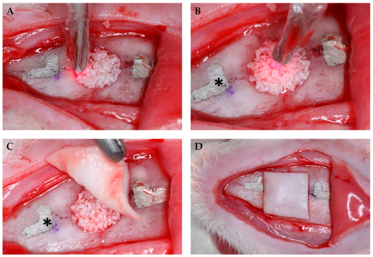
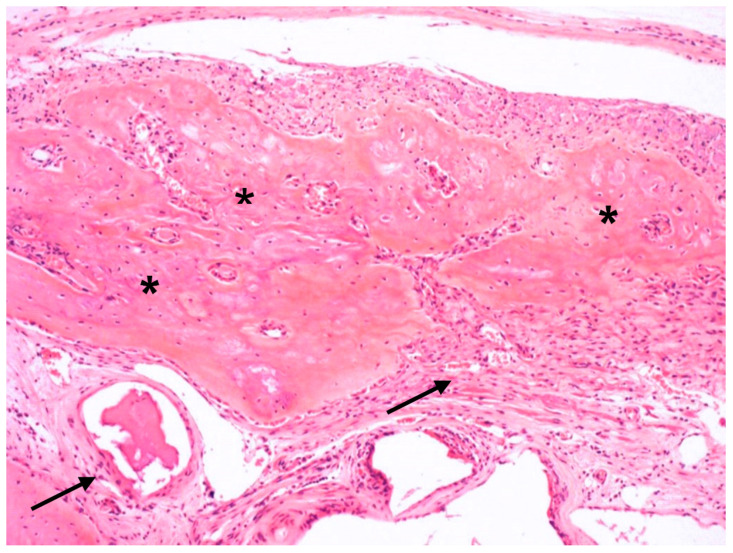
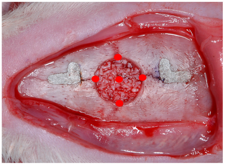
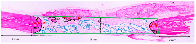
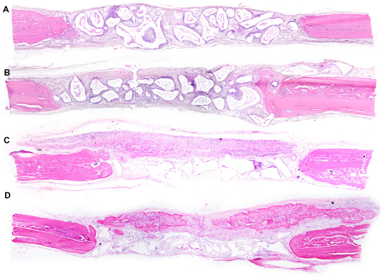
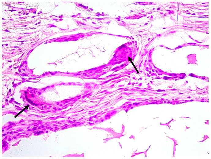
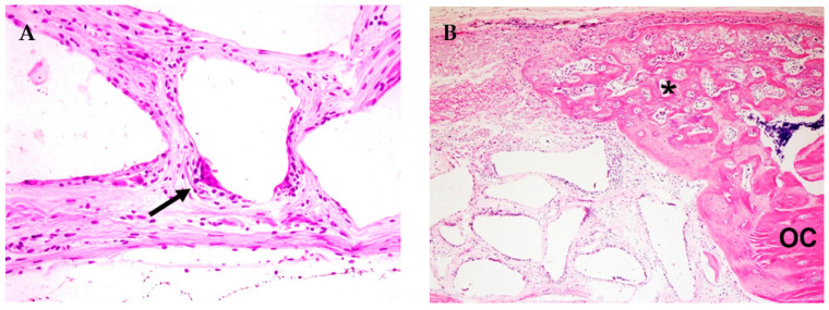
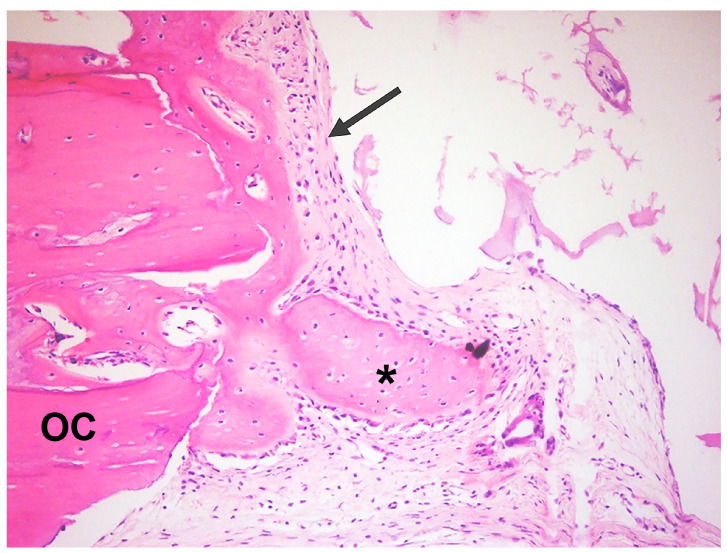
The objective of this study was to evaluate the efficacy of photobiomodulation in the bone regeneration of critical-sized defects (CSD) filled with inorganic bovine bone associated or not with collagen membranes. The study has been conducted on 40 critical defects in the calvaria of male rats, divided into four experimental groups ( = 10): (1) DBBM (deproteinized bovine bone mineral); (2) GBR (DBBM+collagen membrane); (3) DBBM+P (DBBM+photobiomodulation); and (4) GBR+P (GBR+photobiomodulation). At 30 days postoperative, the animals were euthanized, and after the tissue had been processed, histological, histometric, and statistical analyses were performed. The analyses have taken into account newly formed bone area (NBA), linear bone extension (LBE), and residual particle area (RPA) as variables. The Kruskal-Wallis test has been performed, followed by the Dwass-Steel-Critchlow-Fligner test for comparison between groups ( < 0.05). When the DBBM+P group was compared to the DBBM group, it was possible to observe significant statistical differences in all the variables analyzed ( < 0.05). The application of photobiomodulation in guided bone regeneration (GBR+P) has shown a decrease in the median value for the RPA variable (26.8) when compared to the GBR group (32.4), with a significant statistical difference; however, for NBA and LBE, the therapy has not provided significant results.
本研究的目的是评估光生物调节对填充有无机牛骨且联合或不联合胶原膜的临界尺寸骨缺损(CSD)骨再生的疗效。该研究在雄性大鼠颅骨的40处临界骨缺损上进行,分为四个实验组(每组n = 10):(1)脱蛋白牛骨矿物质(DBBM);(2)引导骨再生(GBR,DBBM + 胶原膜);(3)DBBM + P(DBBM + 光生物调节);以及(4)GBR + P(GBR + 光生物调节)。术后30天,对动物实施安乐死,在对组织进行处理后,进行组织学、组织计量学和统计学分析。分析将新形成骨面积(NBA)、线性骨延伸(LBE)和残余颗粒面积(RPA)作为变量。进行了Kruskal-Wallis检验,随后进行Dwass-Steel-Critchlow-Fligner检验以比较组间差异(P < 0.05)。当将DBBM + P组与DBBM组进行比较时,在所有分析变量中均观察到显著的统计学差异(P < 0.05)。与GBR组(32.4)相比,光生物调节在引导骨再生(GBR + P)中的应用显示RPA变量的中位数有所降低(26.8),具有显著的统计学差异;然而,对于NBA和LBE,该疗法未提供显著结果。