• 文献检索
  • 文档翻译
  • 深度研究
  • 学术资讯
  • Suppr Zotero 插件Zotero 插件
  • 邀请有礼
  • 套餐&价格
  • 历史记录
应用&插件
Suppr Zotero 插件Zotero 插件浏览器插件Mac 客户端Windows 客户端微信小程序
定价
高级版会员购买积分包购买API积分包
服务
文献检索文档翻译深度研究API 文档MCP 服务
关于我们
关于 Suppr公司介绍联系我们用户协议隐私条款
关注我们

Suppr 超能文献

核心技术专利:CN118964589B侵权必究
粤ICP备2023148730 号-1Suppr @ 2026

文献检索

告别复杂PubMed语法,用中文像聊天一样搜索,搜遍4000万医学文献。AI智能推荐,让科研检索更轻松。

立即免费搜索

文件翻译

保留排版,准确专业,支持PDF/Word/PPT等文件格式,支持 12+语言互译。

免费翻译文档

深度研究

AI帮你快速写综述,25分钟生成高质量综述,智能提取关键信息,辅助科研写作。

立即免费体验

人类大脑功能的几何约束。

Geometric constraints on human brain function.

机构信息

The Turner Institute for Brain and Mental Health, School of Psychological Sciences and Monash Biomedical Imaging, Monash University, Clayton, Victoria, Australia.

School of Physics, University of Sydney, Camperdown, New South Wales, Australia.

出版信息

Nature. 2023 Jun;618(7965):566-574. doi: 10.1038/s41586-023-06098-1. Epub 2023 May 31.

DOI:10.1038/s41586-023-06098-1
PMID:37258669
原文链接:https://pmc.ncbi.nlm.nih.gov/articles/PMC10266981/
Abstract

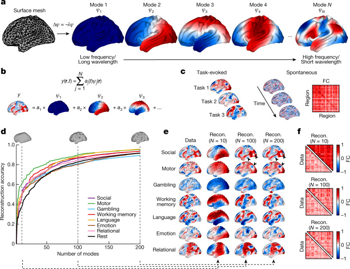
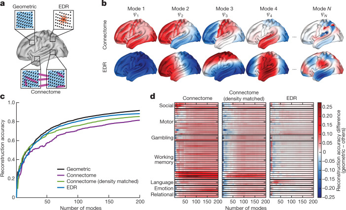
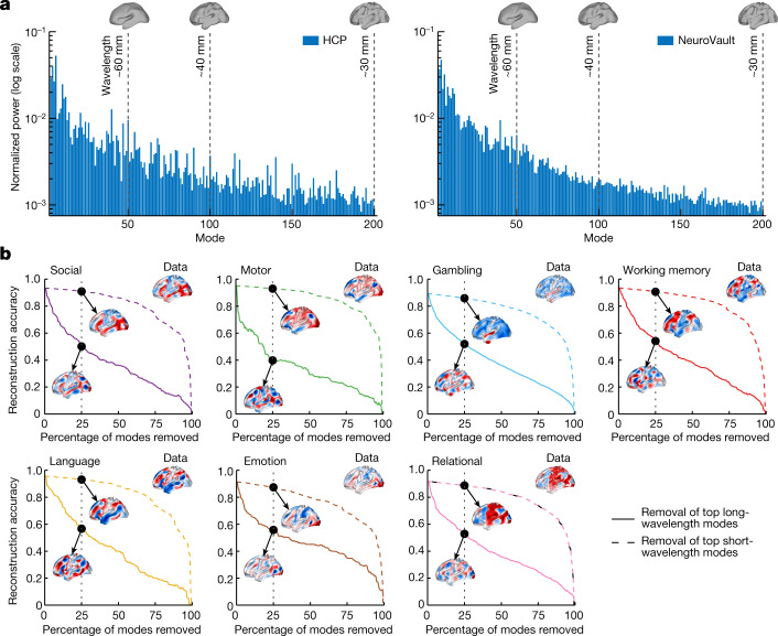
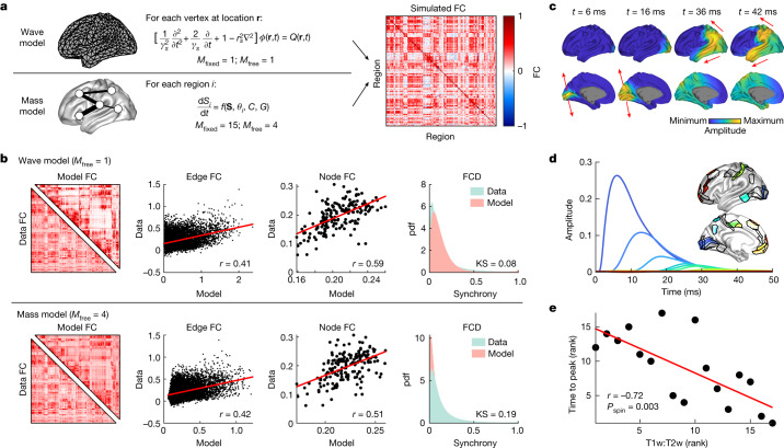
The anatomy of the brain necessarily constrains its function, but precisely how remains unclear. The classical and dominant paradigm in neuroscience is that neuronal dynamics are driven by interactions between discrete, functionally specialized cell populations connected by a complex array of axonal fibres. However, predictions from neural field theory, an established mathematical framework for modelling large-scale brain activity, suggest that the geometry of the brain may represent a more fundamental constraint on dynamics than complex interregional connectivity. Here, we confirm these theoretical predictions by analysing human magnetic resonance imaging data acquired under spontaneous and diverse task-evoked conditions. Specifically, we show that cortical and subcortical activity can be parsimoniously understood as resulting from excitations of fundamental, resonant modes of the brain's geometry (that is, its shape) rather than from modes of complex interregional connectivity, as classically assumed. We then use these geometric modes to show that task-evoked activations across over 10,000 brain maps are not confined to focal areas, as widely believed, but instead excite brain-wide modes with wavelengths spanning over 60 mm. Finally, we confirm predictions that the close link between geometry and function is explained by a dominant role for wave-like activity, showing that wave dynamics can reproduce numerous canonical spatiotemporal properties of spontaneous and evoked recordings. Our findings challenge prevailing views and identify a previously underappreciated role of geometry in shaping function, as predicted by a unifying and physically principled model of brain-wide dynamics.

摘要

大脑的解剖结构必然限制了它的功能,但具体的限制方式仍不清楚。神经科学中的经典和主导范式是,神经元的动力学是由离散的、功能专门化的细胞群体之间的相互作用驱动的,这些细胞群体通过复杂的轴突纤维网络连接。然而,神经场理论的预测,即用于模拟大规模大脑活动的成熟数学框架,表明大脑的几何形状可能是对动力学的更基本限制,而不是复杂的区域间连接。在这里,我们通过分析在自发和多种任务诱发条件下采集的人类磁共振成像数据,证实了这些理论预测。具体来说,我们表明皮质和皮质下的活动可以被简化地理解为是大脑几何形状(即形状)的基本共振模式的激发,而不是经典假设的复杂区域间连接模式的激发。然后,我们使用这些几何模式来表明,跨越超过 10000 个大脑图谱的任务诱发激活并不局限于焦点区域,这与广泛的观点相反,而是激发了具有跨越 60mm 以上波长的全脑模式。最后,我们证实了预测,即几何形状和功能之间的紧密联系是由主导的波动活动解释的,表明波动动力学可以再现自发和诱发记录的许多典型时空特性。我们的发现挑战了现有的观点,并确定了以前被低估的几何形状在塑造功能方面的作用,这与大脑整体动力学的统一和物理原则模型的预测一致。

https://cdn.ncbi.nlm.nih.gov/pmc/blobs/13b5/10266981/b4f9b5a57da8/41586_2023_6098_Fig15_ESM.jpg
https://cdn.ncbi.nlm.nih.gov/pmc/blobs/13b5/10266981/f18e8daf0f19/41586_2023_6098_Fig1_HTML.jpg
https://cdn.ncbi.nlm.nih.gov/pmc/blobs/13b5/10266981/349d9399397a/41586_2023_6098_Fig2_HTML.jpg
https://cdn.ncbi.nlm.nih.gov/pmc/blobs/13b5/10266981/85ef0eb30600/41586_2023_6098_Fig3_HTML.jpg
https://cdn.ncbi.nlm.nih.gov/pmc/blobs/13b5/10266981/f6a3eb36893f/41586_2023_6098_Fig4_HTML.jpg
https://cdn.ncbi.nlm.nih.gov/pmc/blobs/13b5/10266981/f53d1658ada2/41586_2023_6098_Fig5_HTML.jpg
https://cdn.ncbi.nlm.nih.gov/pmc/blobs/13b5/10266981/fab4e903dac5/41586_2023_6098_Fig6_ESM.jpg
https://cdn.ncbi.nlm.nih.gov/pmc/blobs/13b5/10266981/66868c42bb7d/41586_2023_6098_Fig7_ESM.jpg
https://cdn.ncbi.nlm.nih.gov/pmc/blobs/13b5/10266981/476c0a85ef90/41586_2023_6098_Fig8_ESM.jpg
https://cdn.ncbi.nlm.nih.gov/pmc/blobs/13b5/10266981/b6743a174d58/41586_2023_6098_Fig9_ESM.jpg
https://cdn.ncbi.nlm.nih.gov/pmc/blobs/13b5/10266981/aba1181323f0/41586_2023_6098_Fig10_ESM.jpg
https://cdn.ncbi.nlm.nih.gov/pmc/blobs/13b5/10266981/68e8a744dadf/41586_2023_6098_Fig11_ESM.jpg
https://cdn.ncbi.nlm.nih.gov/pmc/blobs/13b5/10266981/5b219313e775/41586_2023_6098_Fig12_ESM.jpg
https://cdn.ncbi.nlm.nih.gov/pmc/blobs/13b5/10266981/5cad665b2c42/41586_2023_6098_Fig13_ESM.jpg
https://cdn.ncbi.nlm.nih.gov/pmc/blobs/13b5/10266981/1095b359f5da/41586_2023_6098_Fig14_ESM.jpg
https://cdn.ncbi.nlm.nih.gov/pmc/blobs/13b5/10266981/b4f9b5a57da8/41586_2023_6098_Fig15_ESM.jpg

相似文献

1
Geometric constraints on human brain function.人类大脑功能的几何约束。
Nature. 2023 Jun;618(7965):566-574. doi: 10.1038/s41586-023-06098-1. Epub 2023 May 31.
2
Transient neuronal coactivations embedded in globally propagating waves underlie resting-state functional connectivity.嵌入全局传播波中的瞬时神经元共激活是静息态功能连接的基础。
Proc Natl Acad Sci U S A. 2016 Jun 7;113(23):6556-61. doi: 10.1073/pnas.1521299113. Epub 2016 May 16.
3
An Evolutionary Game Theory Model of Spontaneous Brain Functioning.自发脑功能的进化博弈论模型。
Sci Rep. 2017 Nov 22;7(1):15978. doi: 10.1038/s41598-017-15865-w.
4
Investigating the neural basis for fMRI-based functional connectivity in a blocked design: application to interregional correlations and psycho-physiological interactions.在组块设计中研究基于功能磁共振成像的功能连接的神经基础:应用于区域间相关性和心理-生理相互作用。
Magn Reson Imaging. 2008 Jun;26(5):583-93. doi: 10.1016/j.mri.2007.10.011. Epub 2008 Jan 10.
5
Near-Critical Dynamics in Stimulus-Evoked Activity of the Human Brain and Its Relation to Spontaneous Resting-State Activity.人类大脑刺激诱发活动中的近临界动力学及其与静息态自发活动的关系。
J Neurosci. 2015 Oct 14;35(41):13927-42. doi: 10.1523/JNEUROSCI.0477-15.2015.
6
A spatiotemporal complexity architecture of human brain activity.人类大脑活动的时空复杂性结构。
Sci Adv. 2023 Feb 3;9(5):eabq3851. doi: 10.1126/sciadv.abq3851. Epub 2023 Feb 1.
7
Neuroelectrical decomposition of spontaneous brain activity measured with functional magnetic resonance imaging.利用功能磁共振成像测量的自发脑活动的神经电分解
Cereb Cortex. 2014 Nov;24(11):3080-9. doi: 10.1093/cercor/bht164. Epub 2013 Jun 24.
8
Cortex-wide neural dynamics predict behavioral states and provide a neural basis for resting-state dynamic functional connectivity.皮质范围的神经动力学预测行为状态,并为静息状态动态功能连接提供神经基础。
Cell Rep. 2023 Jun 27;42(6):112527. doi: 10.1016/j.celrep.2023.112527. Epub 2023 May 26.
9
The Functional Relevance of Task-State Functional Connectivity.任务态功能连接的功能相关性。
J Neurosci. 2021 Mar 24;41(12):2684-2702. doi: 10.1523/JNEUROSCI.1713-20.2021. Epub 2021 Feb 4.
10
Structural Variability in the Human Brain Reflects Fine-Grained Functional Architecture at the Population Level.人类大脑的结构变异性反映了人群水平上的精细功能架构。
J Neurosci. 2019 Jul 31;39(31):6136-6149. doi: 10.1523/JNEUROSCI.2912-18.2019. Epub 2019 May 31.

引用本文的文献

1
Connectome-constrained ligand-receptor interaction analysis for understanding brain network communication.用于理解脑网络通信的连接体约束配体-受体相互作用分析
Nat Commun. 2025 Sep 2;16(1):8179. doi: 10.1038/s41467-025-63204-9.
2
Practice reshapes the geometry and dynamics of task-tailored representations.实践重塑了任务定制表征的几何结构和动态特性。
Cereb Cortex. 2025 Aug 1;35(8). doi: 10.1093/cercor/bhaf125.
3
The organization of serotonergic fibers in the Pacific angelshark brain: neuroanatomical and supercomputing analyses.

本文引用的文献

1
Can hubs of the human connectome be identified consistently with diffusion MRI?人类连接组的中枢能否通过扩散磁共振成像被一致地识别出来?
Netw Neurosci. 2023 Dec 22;7(4):1326-1350. doi: 10.1162/netn_a_00324. eCollection 2023.
2
Evolutionary shaping of human brain dynamics.人类大脑动力学的进化塑造。
Elife. 2022 Oct 26;11:e80627. doi: 10.7554/eLife.80627.
3
The individuality of shape asymmetries of the human cerebral cortex.人类大脑皮层形状不对称的个体差异。
太平洋扁鲨大脑中5-羟色胺能纤维的组织:神经解剖学和超级计算分析
Front Neurosci. 2025 Aug 8;19:1602116. doi: 10.3389/fnins.2025.1602116. eCollection 2025.
4
The effect of spherical projection on spin tests for brain maps.球面投影对脑图谱自旋测试的影响。
Imaging Neurosci (Camb). 2025 Aug 21;3. doi: 10.1162/IMAG.a.118. eCollection 2025.
5
Generalized learning induced by training and tDCS is predicted by prefrontal cortical morphology.前额叶皮层形态可预测由训练和经颅直流电刺激诱导的广义学习。
Cereb Cortex. 2025 Aug 1;35(8). doi: 10.1093/cercor/bhaf229.
6
Generation of surrogate brain maps preserving spatial autocorrelation through random rotation of geometric eigenmodes.通过几何本征模式的随机旋转生成保留空间自相关的替代脑图谱。
Imaging Neurosci (Camb). 2025 Jul 16;3. doi: 10.1162/IMAG.a.71. eCollection 2025.
7
V2C-Long: Longitudinal cortex reconstruction with spatiotemporal correspondence.V2C-Long:具有时空对应关系的纵向皮质重建。
Imaging Neurosci (Camb). 2025 Mar 7;3. doi: 10.1162/imag_a_00500. eCollection 2025.
8
Evidence for a compensatory relationship between left- and right-lateralized brain networks.左右侧化脑网络之间补偿关系的证据。
Imaging Neurosci (Camb). 2025 Jan 29;3. doi: 10.1162/imag_a_00437. eCollection 2025.
9
Perinatal development of structural thalamocortical connectivity.丘脑皮质结构连接的围产期发育
Imaging Neurosci (Camb). 2025 Jan 8;3. doi: 10.1162/imag_a_00418. eCollection 2025.
10
Structure-function coupling and decoupling during movie watching and resting state: Novel insights bridging EEG and structural imaging.观影与静息状态下的结构-功能耦合和解耦:连接脑电图(EEG)与结构成像的新见解
Imaging Neurosci (Camb). 2025 Jan 23;3. doi: 10.1162/imag_a_00448. eCollection 2025.
Elife. 2022 Oct 5;11:e75056. doi: 10.7554/eLife.75056.
4
A parsimonious description of global functional brain organization in three spatiotemporal patterns.用三个时空模式来简约地描述全球功能大脑组织。
Nat Neurosci. 2022 Aug;25(8):1093-1103. doi: 10.1038/s41593-022-01118-1. Epub 2022 Jul 28.
5
An estimation of the absolute number of axons indicates that human cortical areas are sparsely connected.估计轴突的绝对数量表明,人类皮质区域的连接稀疏。
PLoS Biol. 2022 Mar 14;20(3):e3001575. doi: 10.1371/journal.pbio.3001575. eCollection 2022 Mar.
6
On the intersection between data quality and dynamical modelling of large-scale fMRI signals.在大规模 fMRI 信号的数据质量和动态建模的交叉点上。
Neuroimage. 2022 Aug 1;256:119051. doi: 10.1016/j.neuroimage.2022.119051. Epub 2022 Mar 8.
7
Mapping dopaminergic projections in the human brain with resting-state fMRI.利用静息态 fMRI 技术对人类大脑中的多巴胺能投射进行映射。
Elife. 2022 Feb 3;11:e71846. doi: 10.7554/eLife.71846.
8
The connectome spectrum as a canonical basis for a sparse representation of fast brain activity.连接组谱作为快速脑活动稀疏表示的规范基础。
Neuroimage. 2021 Dec 1;244:118611. doi: 10.1016/j.neuroimage.2021.118611. Epub 2021 Sep 21.
9
Determination of Dynamic Brain Connectivity via Spectral Analysis.通过频谱分析确定动态脑连接性
Front Hum Neurosci. 2021 Jul 16;15:655576. doi: 10.3389/fnhum.2021.655576. eCollection 2021.
10
Global waves synchronize the brain's functional systems with fluctuating arousal.全球波动使大脑功能系统与波动的唤醒状态同步。
Sci Adv. 2021 Jul 21;7(30). doi: 10.1126/sciadv.abf2709. Print 2021 Jul.