Department of Ophthalmology, Ninth People's Hospital Affiliated to Shanghai Jiao Tong University School of Medicine, Shanghai, 200011, China.
Shanghai Key Laboratory of Orbital Diseases and Ocular Oncology, Huangpu District, No. 639 Zhizaoju Road, Shanghai, 200011, China.
BMC Ophthalmol. 2023 Jun 7;23(1):256. doi: 10.1186/s12886-023-03001-4.
To perform an in vivo evaluation of the changes in Schlemm's canal (SC) among patients with primary angle-closure disease (PACD) using swept-source optical coherence tomography (SS-OCT).
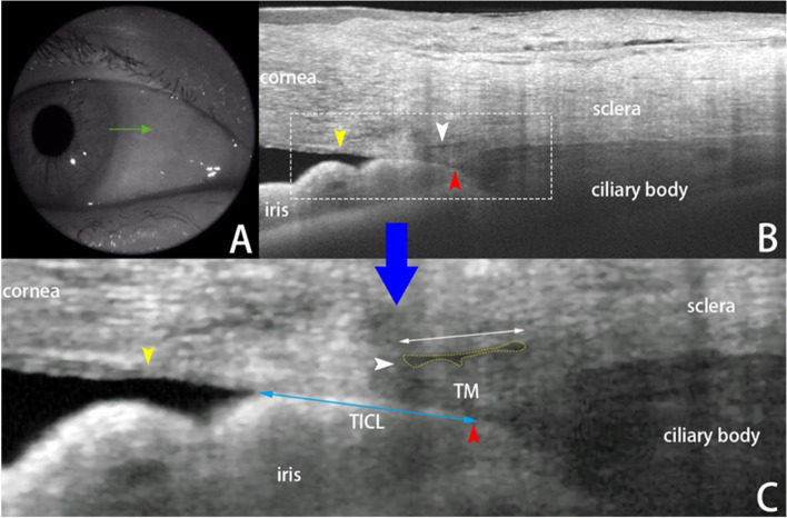
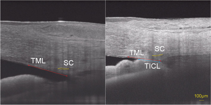
Patients diagnosed with PACD who had not undergone surgery were recruited. The SS-OCT quadrants scanned herein included the nasal and temporal sections at 3 and 9 o'clock, respectively. The diameter and cross-sectional area of the SC were measured. A linear mixed-effects model was performed to analyze the effects of parameters on the SC changes. The hypothesis of interest was related to the angle status (iridotrabecular contact, ITC/open angle, OPN), which was further explored with pairwise comparisons of the estimated marginal means (EMMs) of the SC diameter and SC area. In the ITC regions, the relationship between the trabecular-iris contact length (TICL) percentage and SC parameters was also studied by a mixed model.
A total of 49 eyes of 35 patients were included for measurements and analysis. The percentage of observable SCs in the ITC regions was only 58.5% (24/41), whereas it was 86.0% (49/57) in the OPN regions (χ = 9.44, p = 0.002). ITC was significantly associated with a decreasing SC size. The EMMs for the diameter and cross-sectional area of SC at the ITC and OPN regions were 203.34 μm versus 261.41 μm (p = 0.006) and 3174.43 μm versus 5347.63 μm (p = 0.022), respectively. Sex, age, spherical equivalent refraction, intraocular pressure, axial length, extent of angle closure, history of acute attack and treatment with LPI were not significantly associated with SC parameters. In the ITC regions, a larger TICL percentage was significantly associated with a decrease in SC diameter and area (p = 0.003 and 0.019, respectively).
The morphologies of SC could be affected by the angle status (ITC/OPN) in patients with PACD, and ITC was significantly associated with a decreasing SC size. These changes in SC as described by OCT scans might help to elucidate the progression mechanisms of PACD.
利用扫频源光学相干断层扫描(SS-OCT)对原发性闭角型青光眼(PACG)患者的房水流出道(SC)变化进行体内评估。
招募未接受手术的 PACG 患者。本研究中扫描的 SS-OCT 象限包括 3 点和 9 点的鼻侧和颞侧部分。测量 SC 的直径和横截面积。采用线性混合效应模型分析参数对 SC 变化的影响。感兴趣的假设与角度状态(房角小梁接触、开角、OPN)有关,进一步通过 SC 直径和 SC 面积的估计边际均值(EMM)的两两比较来探讨。在房角小梁接触(TIC)区,还通过混合模型研究了小梁虹膜接触长度(TICL)百分比与 SC 参数之间的关系。
共纳入 35 例 49 只眼进行测量和分析。在房角小梁接触区,可观察到的 SC 百分比仅为 58.5%(24/41),而在开角区为 86.0%(49/57)(χ²=9.44,p=0.002)。房角小梁接触与 SC 变小显著相关。房角小梁接触和开角区 SC 的 EMM 直径和横截面积分别为 203.34μm 比 261.41μm(p=0.006)和 3174.43μm 比 5347.63μm(p=0.022)。性别、年龄、等效球镜屈光度、眼内压、眼轴长度、房角关闭程度、急性发作史和 LPI 治疗与 SC 参数无显著相关性。在房角小梁接触区,较大的 TICL 百分比与 SC 直径和面积减小显著相关(p=0.003 和 0.019)。
PACG 患者的 SC 形态可能受角度状态(房角小梁接触/开角)的影响,房角小梁接触与 SC 变小显著相关。OCT 扫描显示的 SC 变化可能有助于阐明 PACG 的进展机制。