Department of Bioengineering, George Mason University, Fairfax, Virginia, USA.
Center for Computational Fluid Dynamics, College of Science, George Mason University, Fairfax, Virginia, USA.
Int J Numer Method Biomed Eng. 2023 Aug;39(8):e3740. doi: 10.1002/cnm.3740. Epub 2023 Jun 8.
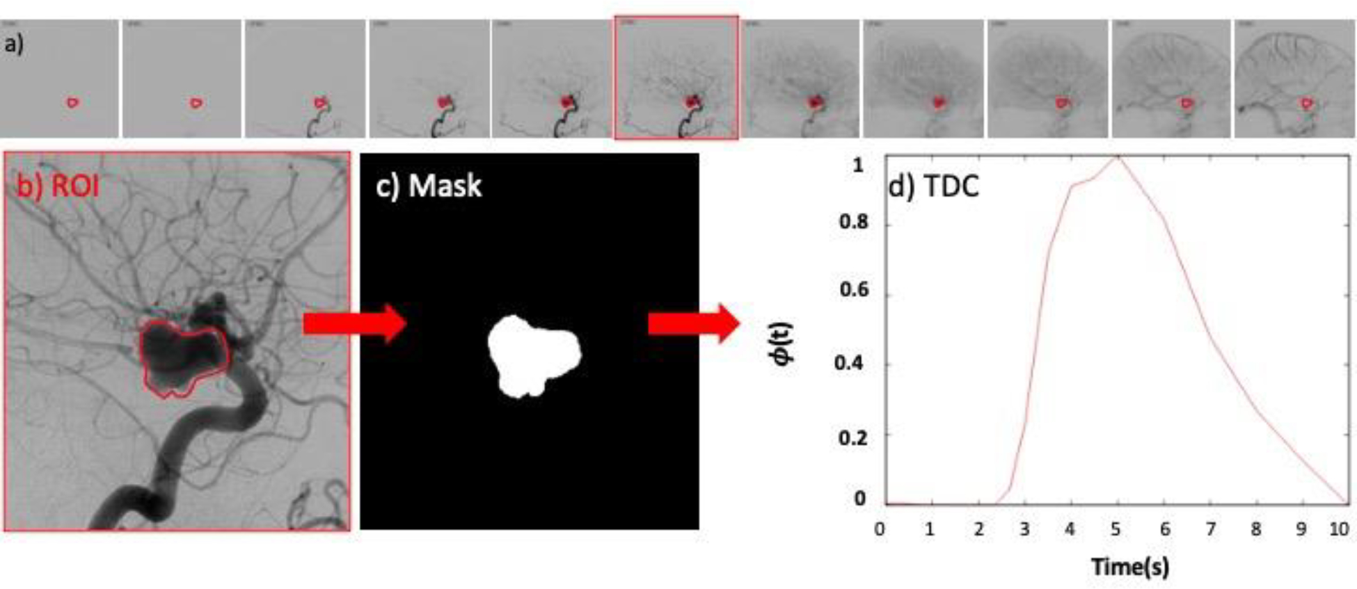
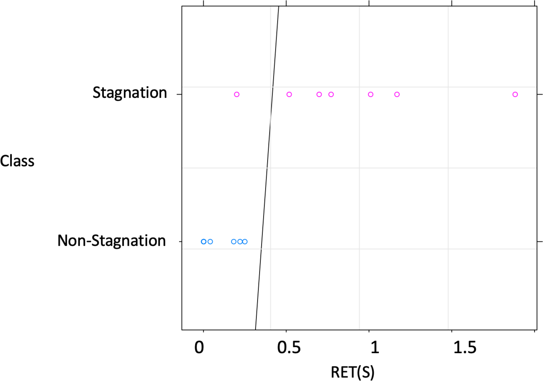
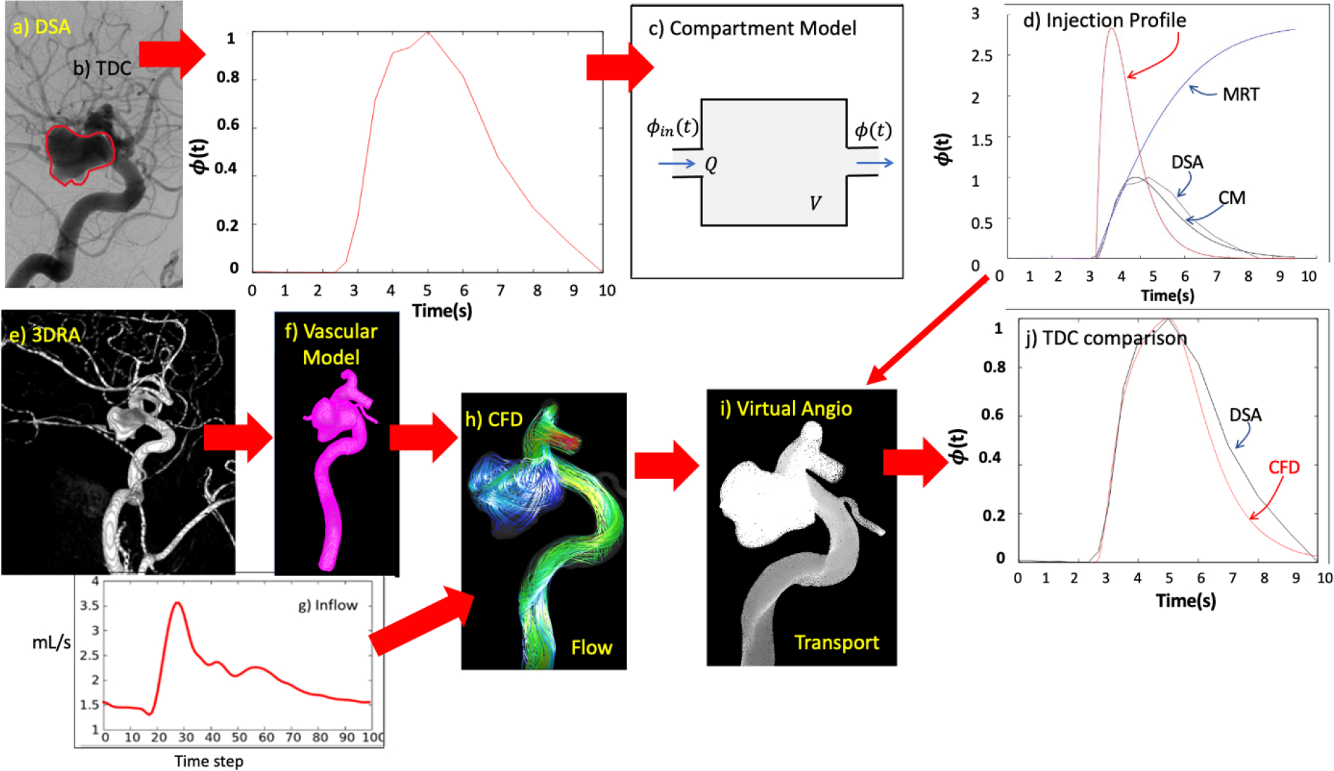
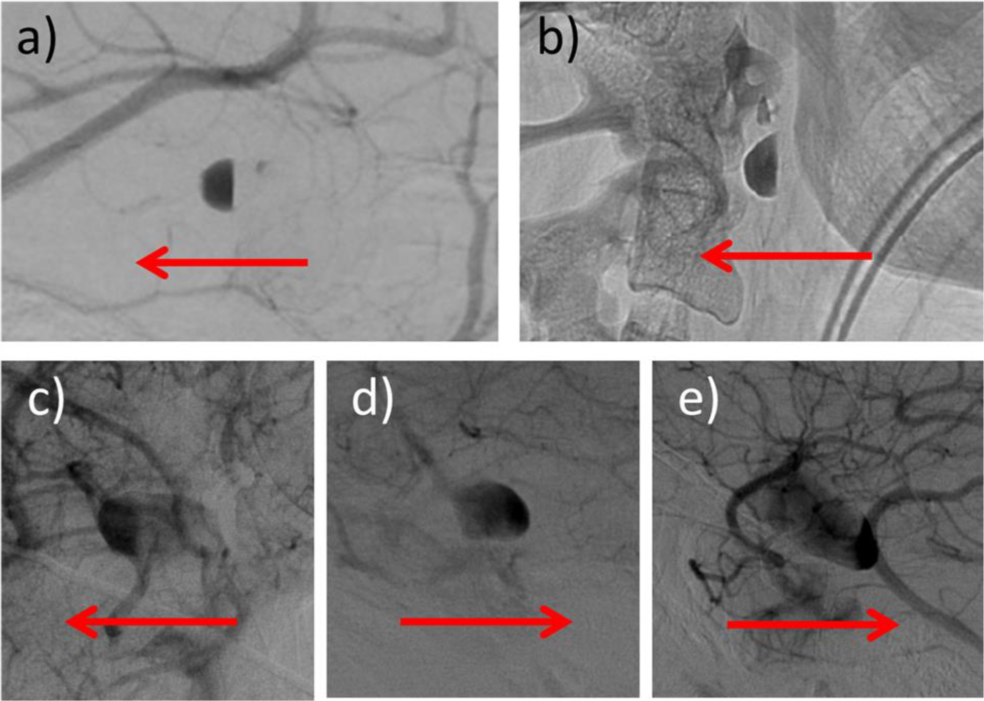
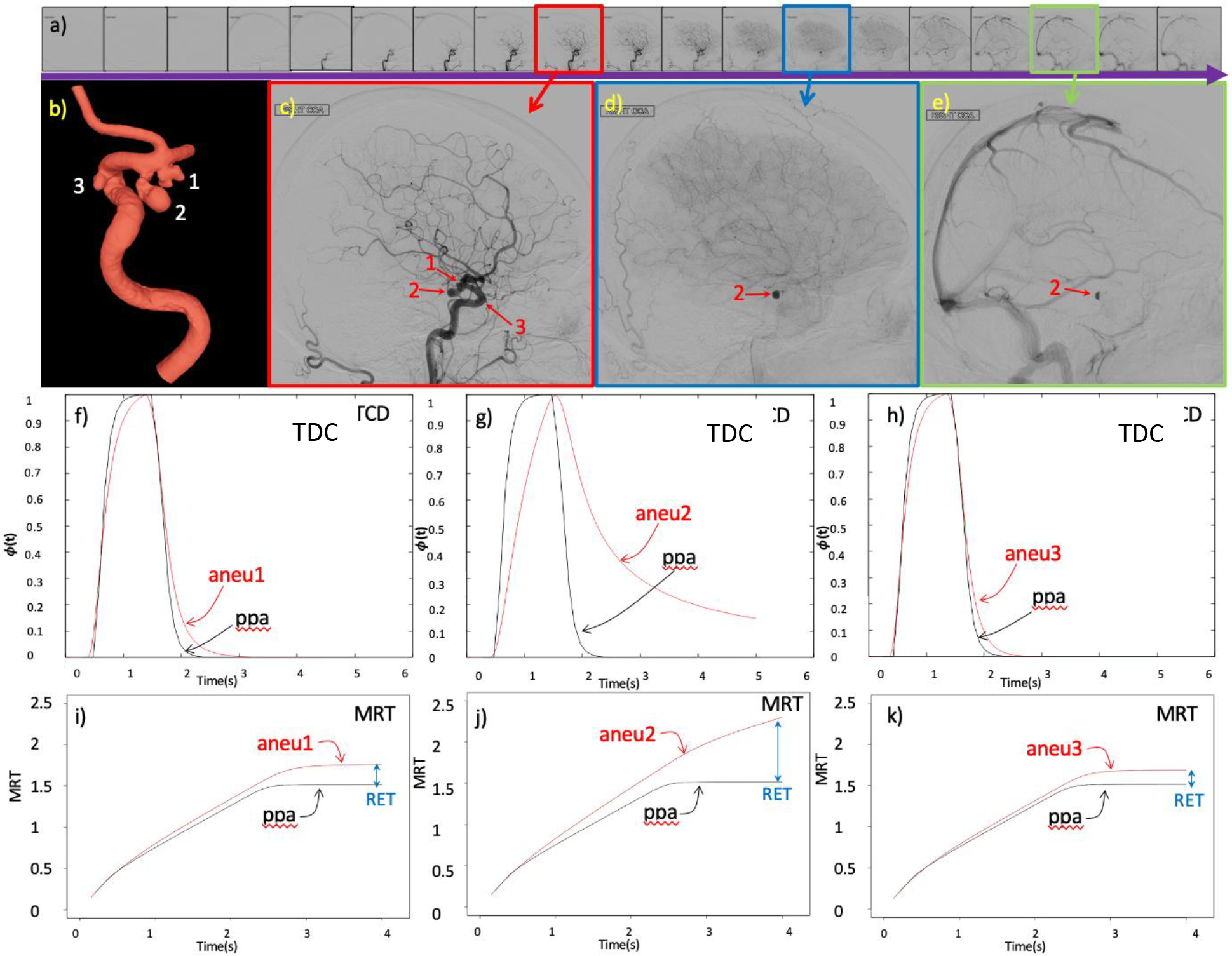
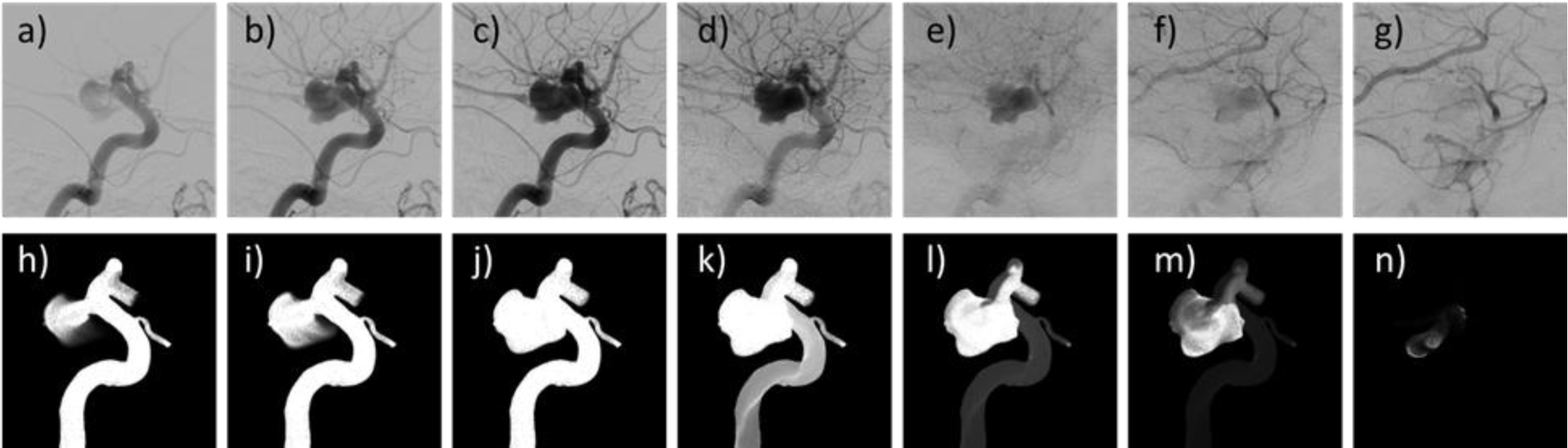
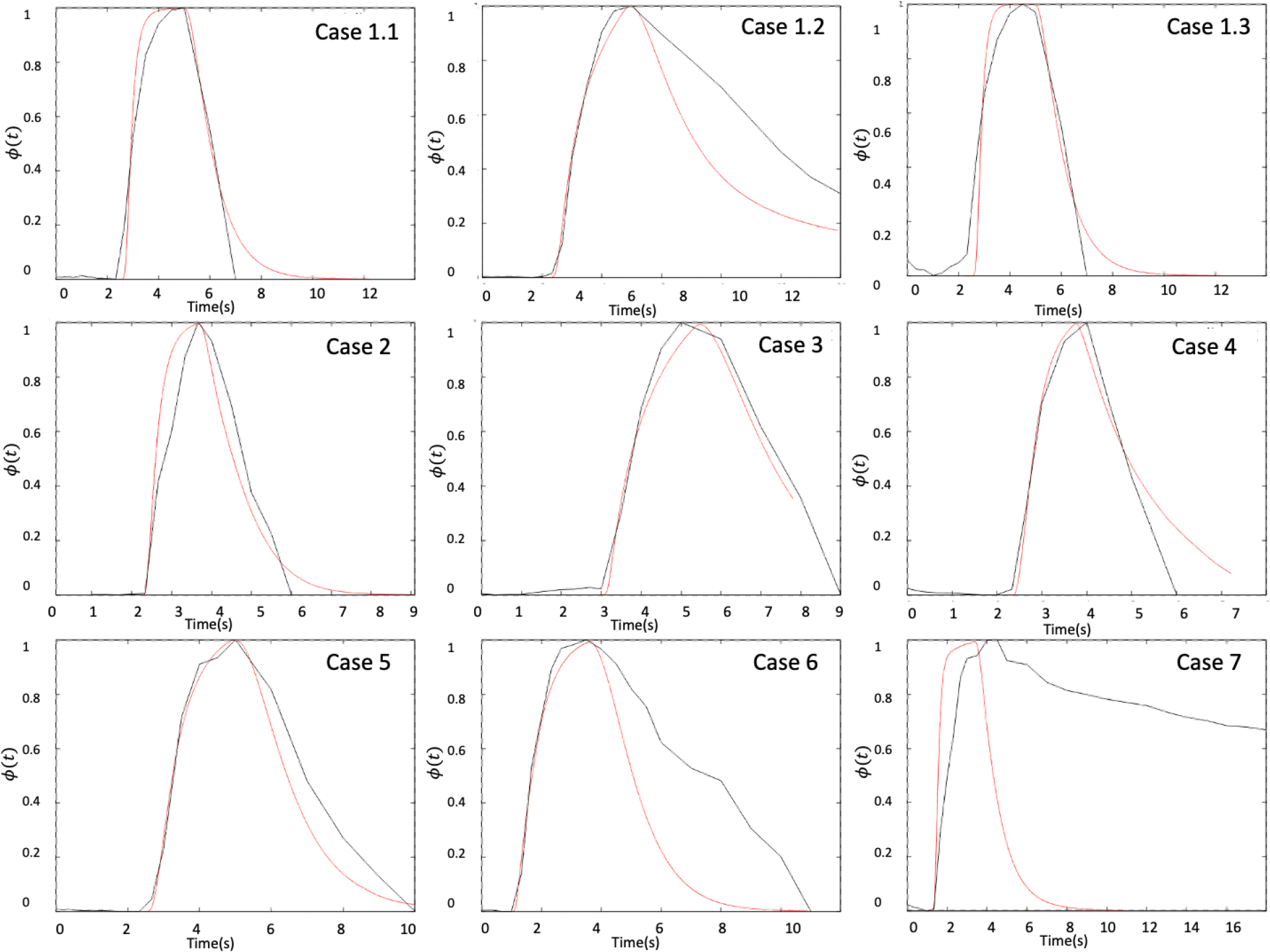
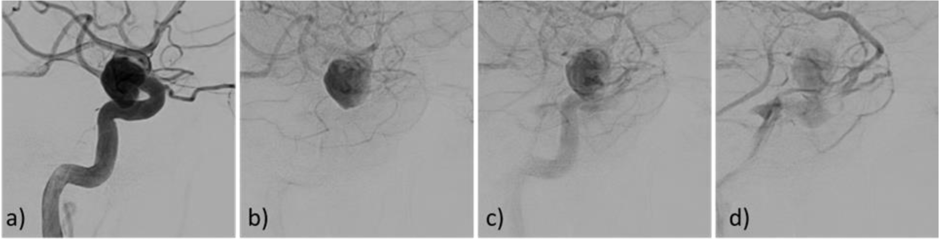
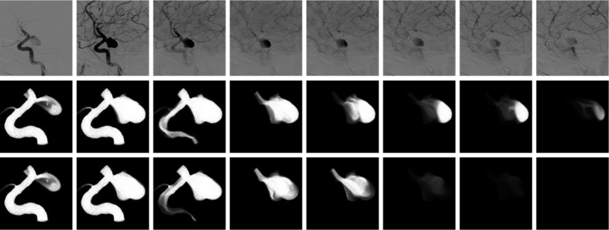
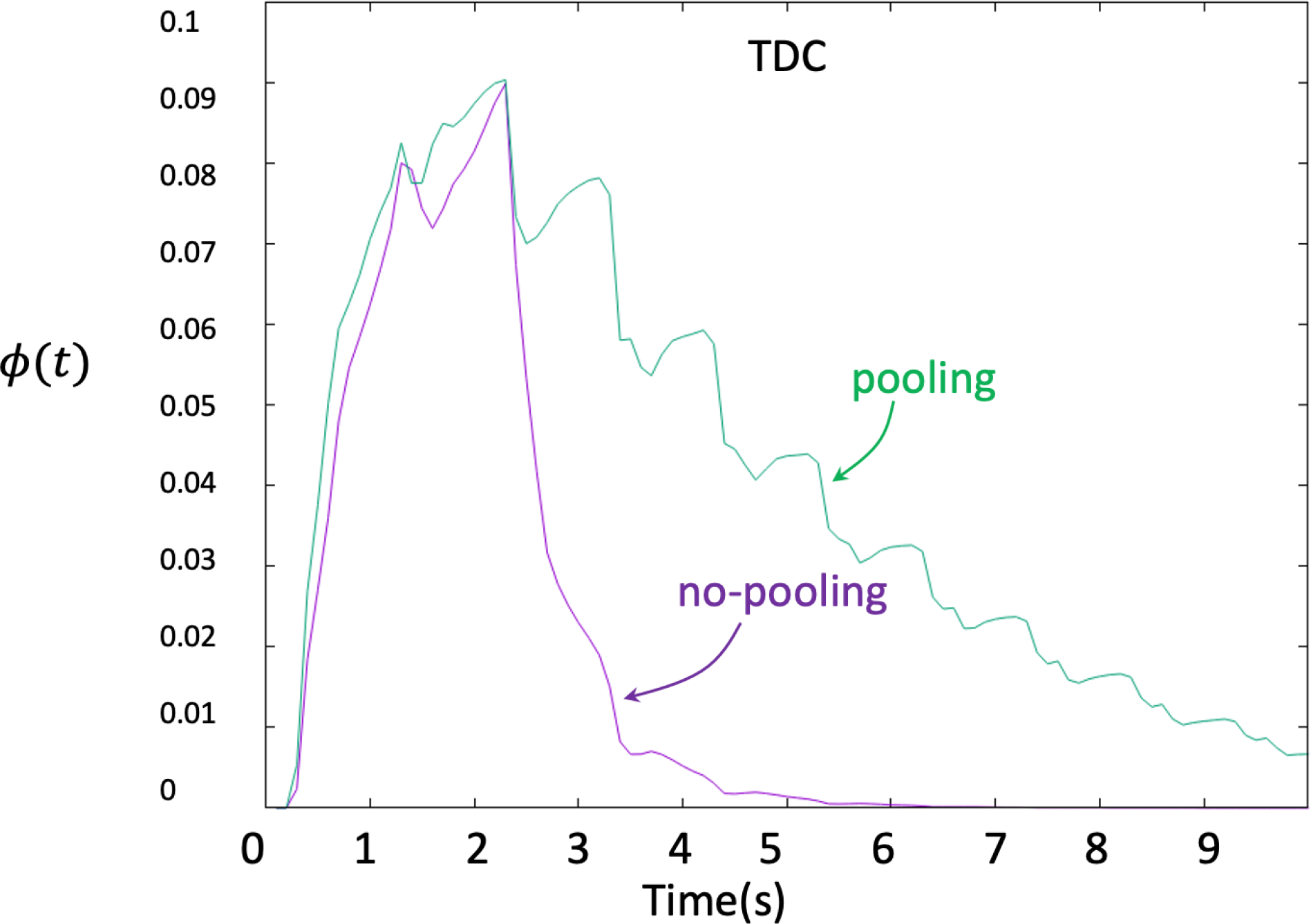
The goal of this study was to test if CFD-based virtual angiograms could be used to automatically discriminate between intracranial aneurysms (IAs) with and without flow stagnation. Time density curves (TDC) were extracted from patient digital subtraction angiography (DSA) image sequences by computing the average gray level intensity inside the aneurysm region and used to define injection profiles for each subject. Subject-specific 3D models were reconstructed from 3D rotational angiography (3DRA) and computational fluid dynamics (CFD) simulations were performed to simulate the blood flow inside IAs. Transport equations were solved numerically to simulate the dynamics of contrast injection into the parent arteries and IAs and then the contrast retention time (RET) was calculated. The importance of gravitational pooling of contrast agent within the aneurysm was evaluated by modeling contrast agent and blood as a mixture of two fluids with different densities and viscosities. Virtual angiograms can reproduce DSA sequences if the correct injection profile is used. RET can identify aneurysms with significant flow stagnation even when the injection profile is not known. Using a small sample of 14 IAs of which seven were previously classified as having flow stagnation, it was found that a threshold RET value of 0.46 s can successfully identify flow stagnation. CFD-based prediction of stagnation was in more than 90% agreement with independent visual DSA assessment of stagnation in a second sample of 34 IAs. While gravitational pooling prolonged contrast retention time it did not affect the predictive capabilities of RET. CFD-based virtual angiograms can detect flow stagnation in IAs and can be used to automatically identify aneurysms with flow stagnation even without including gravitational effects on contrast agents.
本研究旨在测试基于计算流体动力学(CFD)的虚拟血管造影是否可用于自动区分是否存在血流停滞的颅内动脉瘤(IA)。通过计算动脉瘤区域内的平均灰度强度,从患者的数字减影血管造影(DSA)图像序列中提取时间密度曲线(TDC),并用于为每个受试者定义注射曲线。从 3D 旋转血管造影(3DRA)重建受试者特定的 3D 模型,并进行 CFD 模拟以模拟 IA 内的血流。通过数值求解传输方程来模拟对比剂注入到母动脉和 IA 内的动力学,然后计算对比剂保留时间(RET)。通过将对比剂和血液建模为两种具有不同密度和粘度的流体混合物,评估了对比剂在动脉瘤内的重力聚积的重要性。如果使用正确的注射曲线,虚拟血管造影可以再现 DSA 序列。即使不知道注射曲线,RET 也可以识别存在明显血流停滞的动脉瘤。使用先前分类为存在血流停滞的 7 个的 14 个 IA 的小样本,发现 0.46s 的 RET 值可以成功识别血流停滞。在第二个包含 34 个 IA 的样本中,使用 CFD 预测的停滞与独立的视觉 DSA 评估停滞的吻合率超过 90%。虽然重力聚积会延长对比剂保留时间,但它不会影响 RET 的预测能力。基于 CFD 的虚拟血管造影可以检测 IA 中的血流停滞,并可用于自动识别存在血流停滞的动脉瘤,即使不包括对比剂的重力效应。