Department of Dermatology, University of Tübingen, Liebermeisterstr. 25, 72076 Tübingen, Germany; Cluster of Excellence iFIT (EXC 2180) "Image-Guided and Functionally Instructed Tumor Therapies", Tübingen, Germany.
Department of Dermatology, University of Heidelberg, Im Neuenheimer Feld 440, 69120 Heidelberg, Germany.
EBioMedicine. 2023 Jul;93:104644. doi: 10.1016/j.ebiom.2023.104644. Epub 2023 Jun 7.
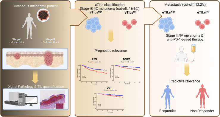
Recent advances in digital pathology have enabled accurate and standardised enumeration of tumour-infiltrating lymphocytes (TILs). Here, we aim to evaluate TILs as a percentage electronic TIL score (eTILs) and investigate its prognostic and predictive relevance in cutaneous melanoma.
We included stage I to IV cutaneous melanoma patients and used hematoxylin-eosin-stained slides for TIL analysis. We assessed eTILs as a continuous and categorical variable using the published cut-off of 16.6% and applied Cox regression models to evaluate associations of eTILs with relapse-free, distant metastasis-free, and overall survival. We compared eTILs of the primaries with matched metastasis. Moreover, we assessed the predictive relevance of eTILs in therapy-naïve metastases according to the first-line therapy.
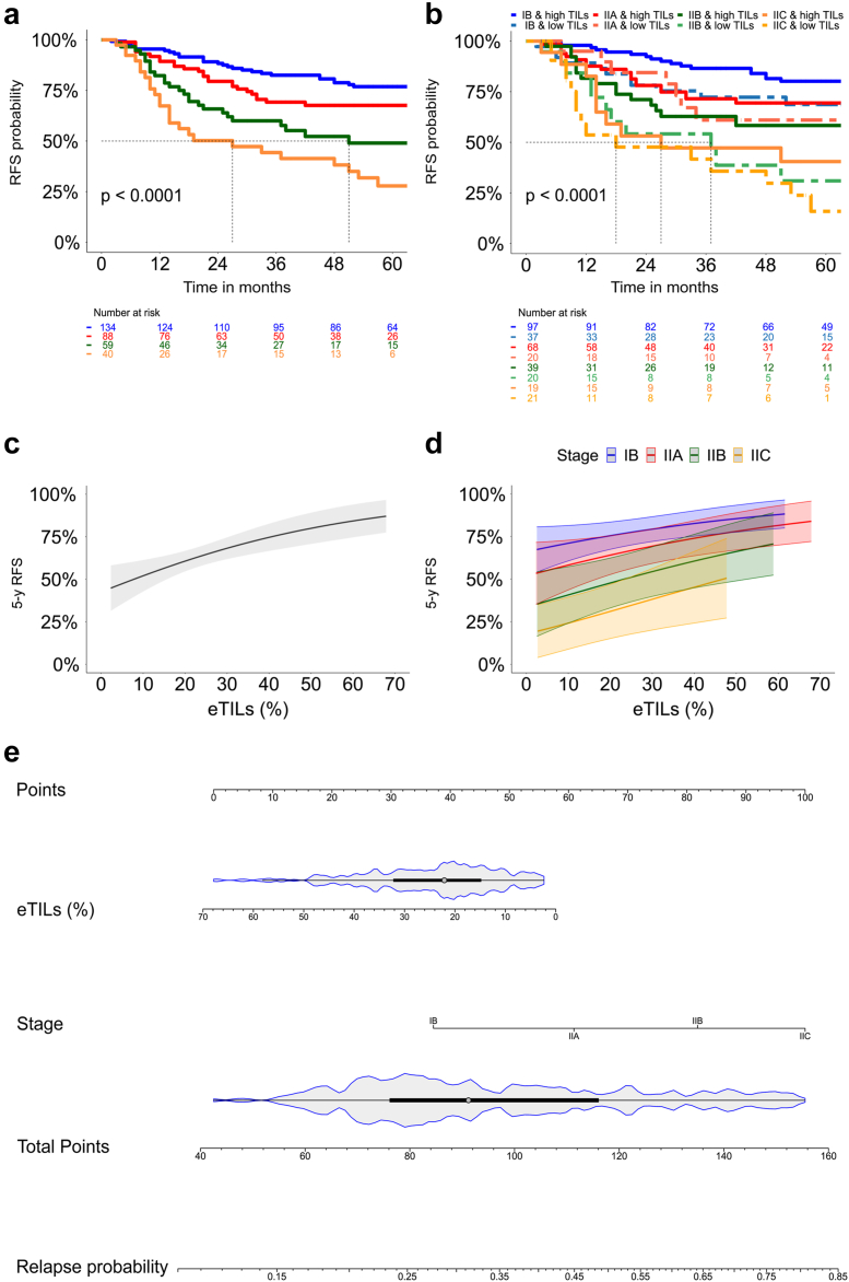
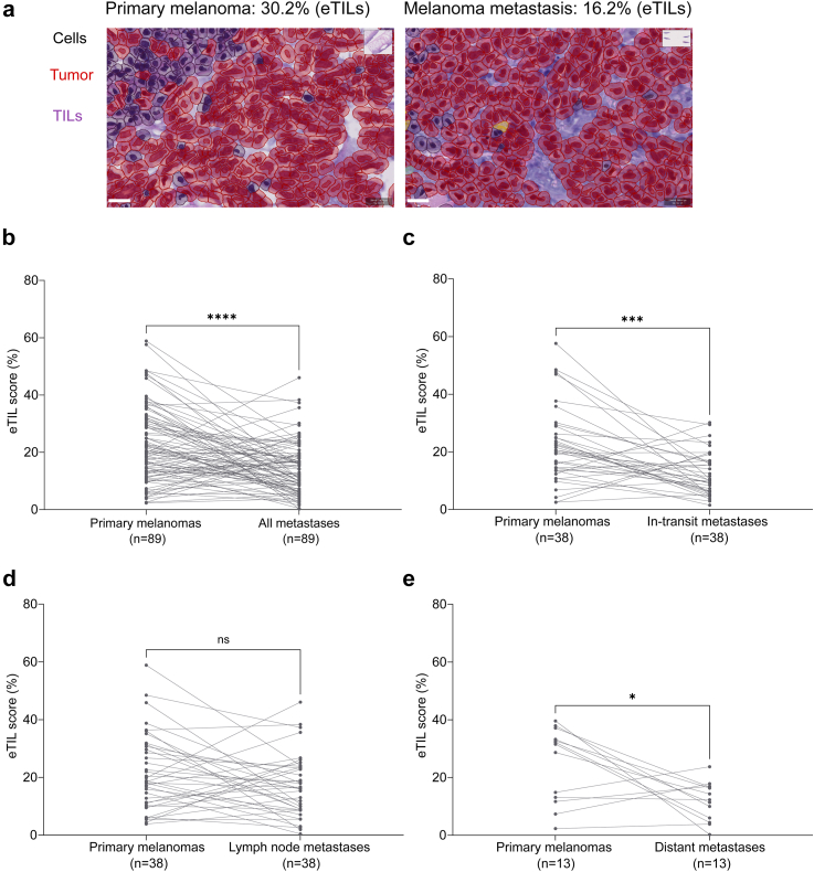
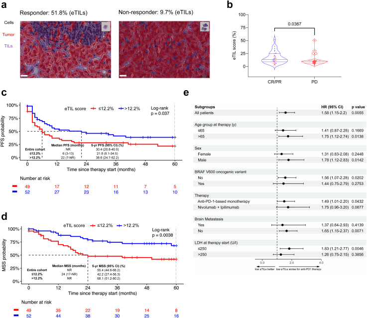
We analysed 321 primary cutaneous melanomas and 191 metastatic samples. In simple Cox regression, tumour thickness (p < 0.0001), presence of ulceration (p = 0.0001) and eTILs ≤16.6% (p = 0.0012) were found to be significant unfavourable prognostic factors for RFS. In multiple Cox regression, eTILs ≤16.6% (p = 0.0161) remained significant and downgraded the current staging. Lower eTILs in the primary tissue was associated with unfavourable relapse-free (p = 0.0014) and distant metastasis-free survival (p = 0.0056). In multiple Cox regression adjusted for tumour thickness and ulceration, eTILs as continuous remained significant (p = 0.019). When comparing TILs in primary tissue and corresponding metastasis of the same patient, eTILs in metastases was lower than in primary melanomas (p < 0.0001). In therapy-naïve metastases, an eTILs >12.2% was associated with longer progression-free survival (p = 0.037) and melanoma-specific survival (p = 0.0038) in patients treated with anti-PD-1-based immunotherapy. In multiple Cox regression, lactate dehydrogenase (p < 0.0001) and eTILs ≤12.2% (p = 0.0130) were significantly associated with unfavourable melanoma-specific survival.
Assessment of TILs is prognostic in primary melanoma samples, and the eTILs complements staging. In therapy-naïve metastases, eTILs ≤12.2% is predictive of unfavourable survival outcomes in patients receiving anti-PD-1-based therapy.
See a detailed list of funding bodies in the Acknowledgements section at the end of the manuscript.
数字病理学的最新进展使得肿瘤浸润淋巴细胞(TILs)的准确和标准化计数成为可能。在这里,我们旨在评估 TILs 作为百分比电子 TIL 评分(eTILs),并研究其在皮肤黑色素瘤中的预后和预测相关性。
我们纳入了 I 期至 IV 期皮肤黑色素瘤患者,并使用苏木精-伊红染色切片进行 TIL 分析。我们使用已发表的 16.6%的截断值评估 eTILs 作为连续和分类变量,并应用 Cox 回归模型来评估 eTILs 与无复发生存、无远处转移生存和总生存之间的关联。我们将原发肿瘤的 eTILs 与匹配的转移瘤进行比较。此外,我们根据一线治疗评估了 eTILs 在未经治疗的转移瘤中的预测相关性。
我们分析了 321 例原发性皮肤黑色素瘤和 191 例转移性样本。在简单的 Cox 回归中,肿瘤厚度(p<0.0001)、溃疡存在(p=0.0001)和 eTILs≤16.6%(p=0.0012)被发现是 RFS 的显著不利预后因素。在多变量 Cox 回归中,eTILs≤16.6%(p=0.0161)仍然具有显著意义,并降低了当前的分期。原发组织中较低的 eTILs 与无复发生存(p=0.0014)和无远处转移生存(p=0.0056)不良相关。在调整肿瘤厚度和溃疡的多变量 Cox 回归中,eTILs 作为连续变量仍然具有显著意义(p=0.019)。当比较同一患者的原发组织和相应转移组织中的 TILs 时,转移组织中的 eTILs 低于原发黑色素瘤(p<0.0001)。在未经治疗的转移瘤中,eTILs>12.2%与接受抗 PD-1 为基础的免疫治疗的患者的无进展生存期(p=0.037)和黑色素瘤特异性生存期(p=0.0038)较长相关。在多变量 Cox 回归中,乳酸脱氢酶(p<0.0001)和 eTILs≤12.2%(p=0.0130)与黑色素瘤特异性生存不良显著相关。
TILs 在原发性黑色素瘤样本中的评估具有预后意义,eTILs 补充了分期。在未经治疗的转移瘤中,eTILs≤12.2%是接受抗 PD-1 为基础的治疗的患者不良生存结局的预测指标。
请在文章末尾的致谢部分查看详细的资助机构列表。