• 文献检索
  • 文档翻译
  • 深度研究
  • 学术资讯
  • Suppr Zotero 插件Zotero 插件
  • 邀请有礼
  • 套餐&价格
  • 历史记录
应用&插件
Suppr Zotero 插件Zotero 插件浏览器插件Mac 客户端Windows 客户端微信小程序
定价
高级版会员购买积分包购买API积分包
服务
文献检索文档翻译深度研究API 文档MCP 服务
关于我们
关于 Suppr公司介绍联系我们用户协议隐私条款
关注我们

Suppr 超能文献

核心技术专利:CN118964589B侵权必究
粤ICP备2023148730 号-1Suppr @ 2026

文献检索

告别复杂PubMed语法,用中文像聊天一样搜索,搜遍4000万医学文献。AI智能推荐,让科研检索更轻松。

立即免费搜索

文件翻译

保留排版,准确专业,支持PDF/Word/PPT等文件格式,支持 12+语言互译。

免费翻译文档

深度研究

AI帮你快速写综述,25分钟生成高质量综述,智能提取关键信息,辅助科研写作。

立即免费体验

葡萄膜黑色素瘤的影像学——当前标准及正在研发的方法

Imaging of Uveal Melanoma-Current Standard and Methods in Development.

作者信息

Solnik Małgorzata, Paduszyńska Natalia, Czarnecka Anna M, Synoradzki Kamil J, Yousef Yacoub A, Chorągiewicz Tomasz, Rejdak Robert, Toro Mario Damiano, Zweifel Sandrine, Dyndor Katarzyna, Fiedorowicz Michał

机构信息

Faculty of Medicine, Medical University of Warsaw, 02-091 Warsaw, Poland.

Department of Soft Tissue/Bone Sarcoma and Melanoma, Maria Sklodowska-Curie National Research Institute of Oncology, 5 Roentgen Str., 02-781 Warsaw, Poland.

出版信息

Cancers (Basel). 2022 Jun 27;14(13):3147. doi: 10.3390/cancers14133147.

DOI:10.3390/cancers14133147
PMID:35804919
原文链接:https://pmc.ncbi.nlm.nih.gov/articles/PMC9265106/
Abstract

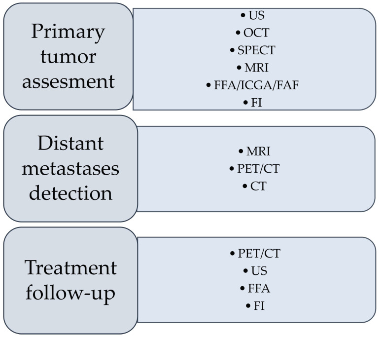
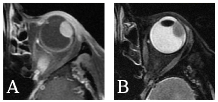
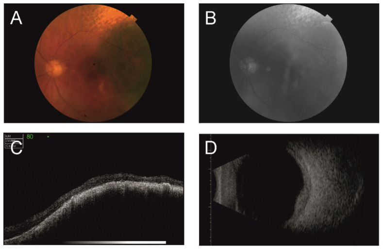
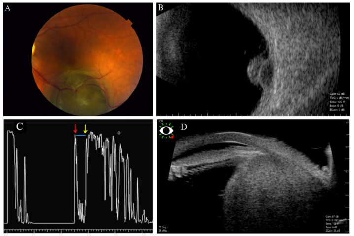
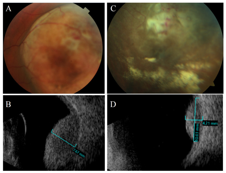
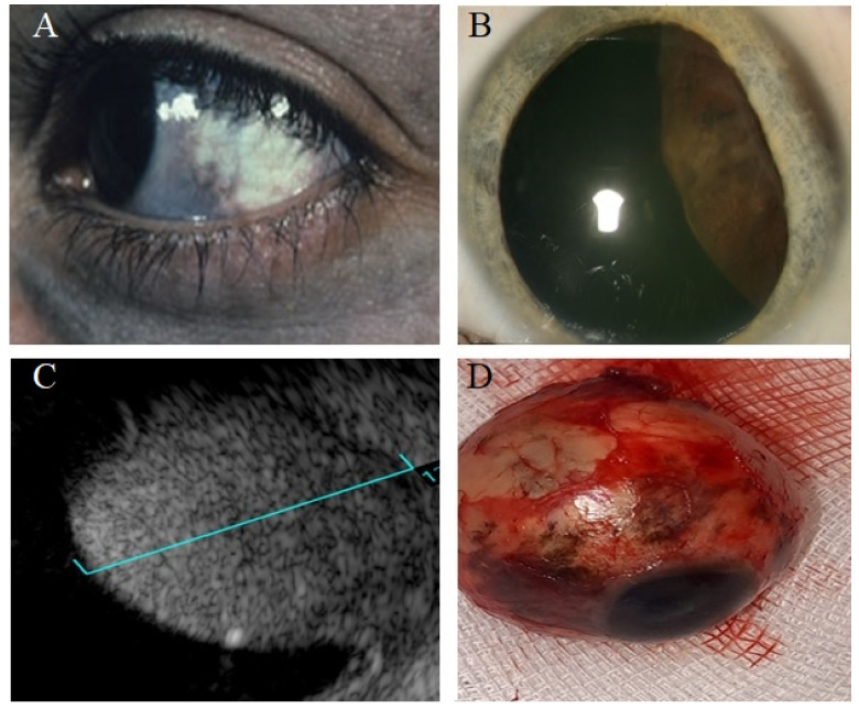
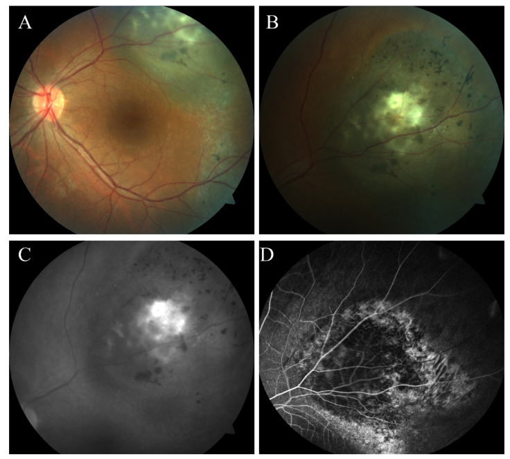
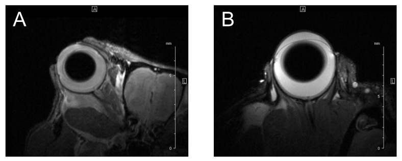
Uveal melanoma is the most common primary intraocular malignancy in adults, characterized by an insidious onset and poor prognosis strongly associated with tumor size and the presence of distant metastases, most commonly in the liver. Contrary to most tumor identification, a biopsy followed by a pathological exam is used only in certain cases. Therefore, an early and noninvasive diagnosis is essential to enhance patients' chances for early treatment. We reviewed imaging modalities currently used in the diagnostics of uveal melanoma, including fundus imaging, ultrasonography (US), optical coherence tomography (OCT), single-photon emission computed tomography (SPECT), fundus fluorescein angiography (FFA), indocyanine green angiography (ICGA), fundus autofluorescence (FAF), as well as positron emission tomography/computed tomography (PET/CT) or magnetic resonance imaging (MRI). The principle of imaging techniques is briefly explained, along with their role in the diagnostic process and a summary of their advantages and limitations. Further, the experimental data and the advancements in imaging modalities are explained. We describe UM imaging innovations, show their current usage and development, and explain the possibilities of utilizing such modalities to diagnose uveal melanoma in the future.

摘要

葡萄膜黑色素瘤是成人最常见的原发性眼内恶性肿瘤,其特点是起病隐匿,预后较差,这与肿瘤大小及远处转移的存在密切相关,最常见的远处转移部位是肝脏。与大多数肿瘤的诊断不同,仅在某些情况下才进行活检及病理检查。因此,早期无创诊断对于提高患者早期治疗的机会至关重要。我们回顾了目前用于葡萄膜黑色素瘤诊断的成像方式,包括眼底成像、超声检查(US)、光学相干断层扫描(OCT)、单光子发射计算机断层扫描(SPECT)、眼底荧光血管造影(FFA)、吲哚菁绿血管造影(ICGA)、眼底自发荧光(FAF),以及正电子发射断层扫描/计算机断层扫描(PET/CT)或磁共振成像(MRI)。简要解释了成像技术的原理,以及它们在诊断过程中的作用,并总结了其优缺点。此外,还阐述了成像方式的实验数据和进展。我们描述了葡萄膜黑色素瘤成像方面的创新,展示了其当前的应用和发展情况,并解释了未来利用这些方式诊断葡萄膜黑色素瘤的可能性。

https://cdn.ncbi.nlm.nih.gov/pmc/blobs/5b96/9265106/06a916fc6f27/cancers-14-03147-g008.jpg
https://cdn.ncbi.nlm.nih.gov/pmc/blobs/5b96/9265106/d73f66daae0d/cancers-14-03147-g001.jpg
https://cdn.ncbi.nlm.nih.gov/pmc/blobs/5b96/9265106/8e2fc2451fe7/cancers-14-03147-g002.jpg
https://cdn.ncbi.nlm.nih.gov/pmc/blobs/5b96/9265106/5ad181d0c685/cancers-14-03147-g003.jpg
https://cdn.ncbi.nlm.nih.gov/pmc/blobs/5b96/9265106/a1b6579be64d/cancers-14-03147-g004.jpg
https://cdn.ncbi.nlm.nih.gov/pmc/blobs/5b96/9265106/e7d8af8d2d5c/cancers-14-03147-g005.jpg
https://cdn.ncbi.nlm.nih.gov/pmc/blobs/5b96/9265106/1a02916f599f/cancers-14-03147-g006.jpg
https://cdn.ncbi.nlm.nih.gov/pmc/blobs/5b96/9265106/6d0b654dabec/cancers-14-03147-g007.jpg
https://cdn.ncbi.nlm.nih.gov/pmc/blobs/5b96/9265106/06a916fc6f27/cancers-14-03147-g008.jpg

相似文献

1
Imaging of Uveal Melanoma-Current Standard and Methods in Development.葡萄膜黑色素瘤的影像学——当前标准及正在研发的方法
Cancers (Basel). 2022 Jun 27;14(13):3147. doi: 10.3390/cancers14133147.
2
Imaging Techniques in the Diagnosis and Management of Ocular Tumors: Prospects and Challenges.眼部肿瘤的诊断和管理中的影像学技术:前景与挑战。
AAPS J. 2018 Sep 5;20(6):97. doi: 10.1208/s12248-018-0259-9.
3
Fundus autofluorescence and spectral-domain optical coherence tomography findings of leopard spots in nanophthalmic uveal effusion syndrome.先天性小眼球葡萄膜渗出综合征中豹斑的眼底自发荧光和频域光相干断层扫描表现。
Graefes Arch Clin Exp Ophthalmol. 2010 Aug;248(8):1199-202. doi: 10.1007/s00417-010-1352-7. Epub 2010 Mar 19.
4
Staging Uveal Melanoma with Whole-Body Positron-Emission Tomography/Computed Tomography and Abdominal Ultrasound: Low Incidence of Metastatic Disease, High Incidence of Second Primary Cancers.采用全身正电子发射断层扫描/计算机断层扫描和腹部超声对葡萄膜黑色素瘤进行分期:转移性疾病发生率低,第二原发性癌症发生率高。
Middle East Afr J Ophthalmol. 2018 Apr-Jun;25(2):91-95. doi: 10.4103/meajo.MEAJO_96_18.
5
(18)F-fluorodeoxyglucose positron emission tomography/computed tomography and magnetic resonance imaging in patients with liver metastases from uveal melanoma: results from a pilot study.(18)F-氟代脱氧葡萄糖正电子发射断层扫描/计算机断层扫描和磁共振成像在眼黑色素瘤肝转移患者中的应用:一项初步研究的结果。
Melanoma Res. 2012 Feb;22(1):63-9. doi: 10.1097/CMR.0b013e32834d3dcb.
6
A Prospective Comparison of F-prostate-specific Membrane Antigen-1007 Positron Emission Tomography Computed Tomography, Whole-body 1.5 T Magnetic Resonance Imaging with Diffusion-weighted Imaging, and Single-photon Emission Computed Tomography/Computed Tomography with Traditional Imaging in Primary Distant Metastasis Staging of Prostate Cancer (PROSTAGE).F-前列腺特异性膜抗原-1007 正电子发射断层扫描计算机断层扫描、全身 1.5T 磁共振成像弥散加权成像与传统成像单光子发射计算机断层扫描/计算机断层扫描在前列腺癌原发远处转移分期(PROSTAGE)中的前瞻性比较
Eur Urol Oncol. 2021 Aug;4(4):635-644. doi: 10.1016/j.euo.2020.06.012. Epub 2020 Jul 13.
7
Importance of PET/CT examination in patients with malignant uveal melanoma.正电子发射断层显像/计算机断层扫描(PET/CT)检查在恶性葡萄膜黑色素瘤患者中的重要性
Cesk Slov Oftalmol. 2020 Winter;76(1):37-44. doi: 10.31348/2020/5.
8
Whole-body F-18-fluoro-2-deoxyglucose positron emission tomography/computed tomography imaging in the follow-up of metastatic uveal melanoma.全身 F-18-氟代-2-脱氧葡萄糖正电子发射断层扫描/计算机断层扫描成像在转移性葡萄膜黑色素瘤随访中的应用。
Melanoma Res. 2010 Dec;20(6):511-6. doi: 10.1097/CMR.0b013e3283403d6c.
9
Whole-body 18F FDG positron emission tomography/computed tomography evaluation of patients with uveal metastasis.全身 18F-FDG 正电子发射断层扫描/计算机断层扫描评估眼葡萄膜转移患者。
Am J Ophthalmol. 2012 Apr;153(4):661-8. doi: 10.1016/j.ajo.2011.09.028. Epub 2012 Jan 20.
10
Uveal Melanoma: Current Trends in Diagnosis and Management.葡萄膜黑色素瘤:诊断与治疗的当前趋势
Turk J Ophthalmol. 2016 Jun;46(3):123-137. doi: 10.4274/tjo.37431. Epub 2016 Jun 6.

引用本文的文献

1
Advances in Positron Emission Tomography/Computed Tomography for Diagnosing and Managing Primary Posterior Uveal Melanoma.正电子发射断层扫描/计算机断层扫描在原发性后葡萄膜黑色素瘤诊断和管理中的进展
Med Sci Monit. 2025 Aug 19;31:e949252. doi: 10.12659/MSM.949252.
2
Diagnostic value of F-FDG PET/CT in the follow-up of conjunctival melanoma.F-FDG PET/CT在结膜黑色素瘤随访中的诊断价值
Ann Nucl Med. 2025 Aug 15. doi: 10.1007/s12149-025-02097-6.
3
Choroidal metastasis as the first sign of small cell lung cancer: A case report.脉络膜转移作为小细胞肺癌的首发症状:一例报告

本文引用的文献

1
New Perspectives for Eye-Sparing Treatment Strategies in Primary Uveal Melanoma.原发性葡萄膜黑色素瘤保眼治疗策略的新视角
Cancers (Basel). 2021 Dec 28;14(1):134. doi: 10.3390/cancers14010134.
2
Prognostic Biomarkers in Uveal Melanoma: The Status Quo, Recent Advances and Future Directions.葡萄膜黑色素瘤的预后生物标志物:现状、最新进展与未来方向
Cancers (Basel). 2021 Dec 25;14(1):96. doi: 10.3390/cancers14010096.
3
Overall Survival Benefit with Tebentafusp in Metastatic Uveal Melanoma.特普替尼治疗转移性葡萄膜黑色素瘤的总生存获益。
Oncol Lett. 2025 Jul 18;30(4):451. doi: 10.3892/ol.2025.15197. eCollection 2025 Oct.
4
A nomogram to predict cancer-specific survival of ocular melanoma.一种预测眼黑色素瘤癌症特异性生存率的列线图。
Discov Oncol. 2025 Jun 12;16(1):1078. doi: 10.1007/s12672-025-02808-5.
5
Current Treatment of Uveal Melanoma.葡萄膜黑色素瘤的当前治疗方法
Cancers (Basel). 2025 Apr 23;17(9):1403. doi: 10.3390/cancers17091403.
6
Yield of Multimodal Imaging in Iris Amelanotic Lesions: A Masked Case-Control Study.多模态成像在无色素性虹膜病变中的诊断价值:一项设盲病例对照研究。
Diseases. 2025 Mar 28;13(4):99. doi: 10.3390/diseases13040099.
7
Estimating uveal melanoma volume with ellipsoid tumour models.使用椭球体肿瘤模型估计葡萄膜黑色素瘤体积。
Acta Ophthalmol. 2025 Sep;103(6):691-698. doi: 10.1111/aos.17492. Epub 2025 Apr 3.
8
Evaluation of OPTOS wide-field fundus image projections for radiotherapy planning of uveal melanoma.用于葡萄膜黑色素瘤放射治疗计划的OPTOS广角眼底图像投影评估
J Appl Clin Med Phys. 2025 Apr;26(4):e70009. doi: 10.1002/acm2.70009. Epub 2025 Feb 17.
9
Differentiating Choroidal Melanomas and Nevi Using a Self-Supervised Deep Learning Model Applied to Clinical Fundoscopy Images.使用应用于临床眼底镜图像的自监督深度学习模型鉴别脉络膜黑色素瘤和痣。
Ophthalmol Sci. 2024 Nov 8;5(2):100647. doi: 10.1016/j.xops.2024.100647. eCollection 2025 Mar-Apr.
10
Nodular Posterior Scleritis Mimicking Non-pigmented Choroidal Melanoma: A Case Report and Literature Review.模仿无色素性脉络膜黑色素瘤的结节状后巩膜炎:一例报告及文献复习
Cureus. 2024 Nov 6;16(11):e73173. doi: 10.7759/cureus.73173. eCollection 2024 Nov.
N Engl J Med. 2021 Sep 23;385(13):1196-1206. doi: 10.1056/NEJMoa2103485.
4
RETINAL MICROVASCULAR CHANGES IN UVEAL MELANOMA FOLLOWING CONBERCEPT INJECTION AFTER PLAQUE RADIOTHERAPY AS DETECTED BY OPTICAL COHERENCE TOMOGRAPHY ANGIOGRAPHY.经光学相干断层扫描血管造影检测,在巩膜放疗后注射康柏西普的葡萄膜黑色素瘤患者中的视网膜微血管变化
Retina. 2021 Dec 1;41(12):2605-2611. doi: 10.1097/IAE.0000000000003236.
5
Can gray values be converted to Hounsfield units? A systematic review.灰度值可否转换为亨氏单位?系统评价。
Dentomaxillofac Radiol. 2022 Jan 1;51(1):20210140. doi: 10.1259/dmfr.20210140. Epub 2021 Jun 19.
6
Evaluating Retinal and Choroidal Perfusion Changes after Isometric and Dynamic Activity Using Optical Coherence Tomography Angiography.使用光学相干断层扫描血管造影术评估等长运动和动态运动后视网膜和脉络膜灌注变化
Diagnostics (Basel). 2021 Apr 29;11(5):808. doi: 10.3390/diagnostics11050808.
7
Current molecular and clinical insights into uveal melanoma (Review).当前对葡萄膜黑色素瘤的分子和临床认识(综述)。
Int J Oncol. 2021 Apr;58(4). doi: 10.3892/ijo.2021.5190. Epub 2021 Mar 2.
8
Application of Multimodal and Molecular Imaging Techniques in the Detection of Choroidal Melanomas.多模态和分子成像技术在脉络膜黑色素瘤检测中的应用
Front Oncol. 2021 Feb 1;10:617868. doi: 10.3389/fonc.2020.617868. eCollection 2020.
9
White matter: A good reference for the signal intensity evaluation in magnetic resonance imaging for the diagnosis of uveal melanoma.脑白质:葡萄膜黑色素瘤磁共振成像信号强度评估的良好参考。
Neuroradiol J. 2021 Apr;34(2):113-119. doi: 10.1177/1971400920973407. Epub 2020 Dec 2.
10
Automated quantitative OCTA measurements of uveal melanoma-treated eyes with and without radiation retinopathy after I-125 plaque therapy and of nonirradiated fellow eyes.采用 I-125 放射性敷贴治疗后伴或不伴放射性视网膜病变的葡萄膜黑色素瘤治疗眼以及未受照射的对侧眼的自动定量 OCTA 测量。
Eye (Lond). 2021 Mar;35(3):769-776. doi: 10.1038/s41433-020-01237-8. Epub 2020 Nov 23.