van den Berg Ingeborg, Savenije Mark H F, Teunissen Frederik R, van de Pol Sandrine M G, Rasing Marnix J A, van Melick Harm H E, Brink Wyger M, de Boer Johannes C J, van den Berg Cornelis A T, van der Voort van Zyp Jochem R N
Department of Radiation Oncology, Division of Imaging & Oncology, University Medical Center Utrecht, Utrecht, The Netherlands.
Department of Urology, St. Antonius Hospital, Nieuwegein, Utrecht, The Netherlands.
Phys Imaging Radiat Oncol. 2023 Jun 1;26:100453. doi: 10.1016/j.phro.2023.100453. eCollection 2023 Apr.
Manual contouring of neurovascular structures on prostate magnetic resonance imaging (MRI) is labor-intensive and prone to considerable interrater disagreement. Our aim is to contour neurovascular structures automatically on prostate MRI by deep learning (DL) to improve workflow and interrater agreement.
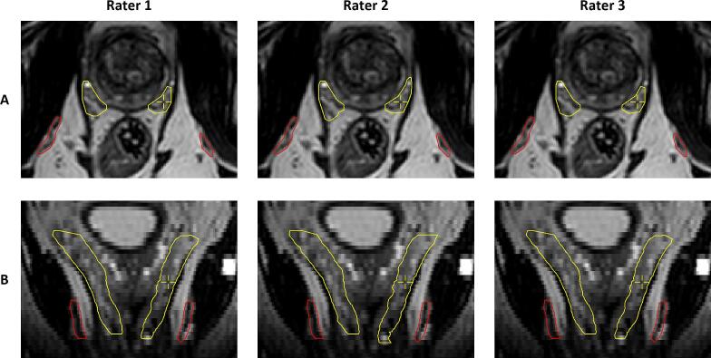
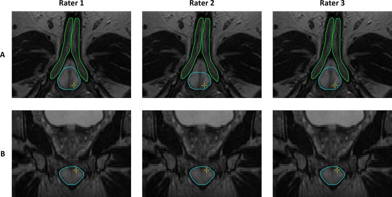
Segmentation of neurovascular structures was performed on pre-treatment 3.0 T MRI data of 131 prostate cancer patients (training [n = 105] and testing [n = 26]). The neurovascular structures include the penile bulb (PB), corpora cavernosa (CCs), internal pudendal arteries (IPAs), and neurovascular bundles (NVBs). Two DL networks, nnU-Net and DeepMedic, were trained for auto-contouring on prostate MRI and evaluated using volumetric Dice similarity coefficient (DSC), mean surface distances (MSD), Hausdorff distances, and surface DSC. Three radiation oncologists evaluated the DL-generated contours and performed corrections when necessary. Interrater agreement was assessed and the time required for manual correction was recorded.
nnU-Net achieved a median DSC of 0.92 (IQR: 0.90-0.93) for the PB, 0.90 (IQR: 0.86-0.92) for the CCs, 0.79 (IQR: 0.77-0.83) for the IPAs, and 0.77 (IQR: 0.72-0.81) for the NVBs, which outperformed DeepMedic for each structure (p < 0.03). nnU-Net showed a median MSD of 0.24 mm for the IPAs and 0.71 mm for the NVBs. The median interrater DSC ranged from 0.93 to 1.00, with the majority of cases (68.9%) requiring manual correction times under two minutes.
DL enables reliable auto-contouring of neurovascular structures on pre-treatment MRI data, easing the clinical workflow in neurovascular-sparing MR-guided radiotherapy.
在前列腺磁共振成像(MRI)上手动勾勒神经血管结构既费力又容易出现评分者间的显著差异。我们的目标是通过深度学习(DL)在前列腺MRI上自动勾勒神经血管结构,以改善工作流程和评分者间的一致性。
对131例前列腺癌患者的治疗前3.0T MRI数据进行神经血管结构分割(训练组[n = 105]和测试组[n = 26])。神经血管结构包括阴茎球(PB)、海绵体(CCs)、阴部内动脉(IPAs)和神经血管束(NVBs)。训练了两个DL网络nnU-Net和DeepMedic,用于在前列腺MRI上自动勾勒轮廓,并使用体积骰子相似系数(DSC)、平均表面距离(MSD)、豪斯多夫距离和表面DSC进行评估。三名放射肿瘤学家评估了DL生成的轮廓,并在必要时进行校正。评估了评分者间的一致性,并记录了手动校正所需的时间。
nnU-Net对PB的DSC中位数为0.92(IQR:0.90 - 0.93),对CCs为0.90(IQR:0.86 - 0.92),对IPAs为0.79(IQR:0.77 - 0.83),对NVBs为0.77(IQR:0.72 - 0.81),在每个结构上均优于DeepMedic(p < 0.03)。nnU-Net对IPAs的MSD中位数为0.24mm,对NVBs为0.71mm。评分者间DSC中位数范围为0.93至1.00,大多数病例(68.9%)手动校正时间在两分钟以内。
DL能够在治疗前MRI数据上可靠地自动勾勒神经血管结构,简化了保留神经血管的MR引导放疗的临床工作流程。