Balagopal Anjali, Dohopolski Michael, Suk Kwon Young, Montalvo Steven, Morgan Howard, Bai Ti, Nguyen Dan, Liang Xiao, Zhong Xinran, Lin Mu-Han, Desai Neil, Jiang Steve
Medical Artificial Intelligence and Automation (MAIA) Laboratory and Department of Radiation Oncology, University of Texas Southwestern Medical Center, Dallas, TX, USA.
Phys Imaging Radiat Oncol. 2024 Apr 15;30:100577. doi: 10.1016/j.phro.2024.100577. eCollection 2024 Apr.
Radiation-induced erectile dysfunction (RiED) commonly affects prostate cancer patients, prompting clinical trials across institutions to explore dose-sparing to internal-pudendal-arteries (IPA) for preserving sexual potency. IPA, challenging to segment, isn't conventionally considered an organ-at-risk (OAR). This study proposes a deep learning (DL) auto-segmentation model for IPA, using Computed Tomography (CT) and Magnetic Resonance Imaging (MRI) or CT alone to accommodate varied clinical practices.
A total of 86 patients with CT and MRI images and noisy IPA labels were recruited in this study. We split the data into 42/14/30 for model training, testing, and a clinical observer study, respectively. There were three major innovations in this model: 1) we designed an architecture with squeeze-and-excite blocks and modality attention for effective feature extraction and production of accurate segmentation, 2) a novel loss function was used for training the model effectively with noisy labels, and 3) modality dropout strategy was used for making the model capable of segmentation in the absence of MRI.
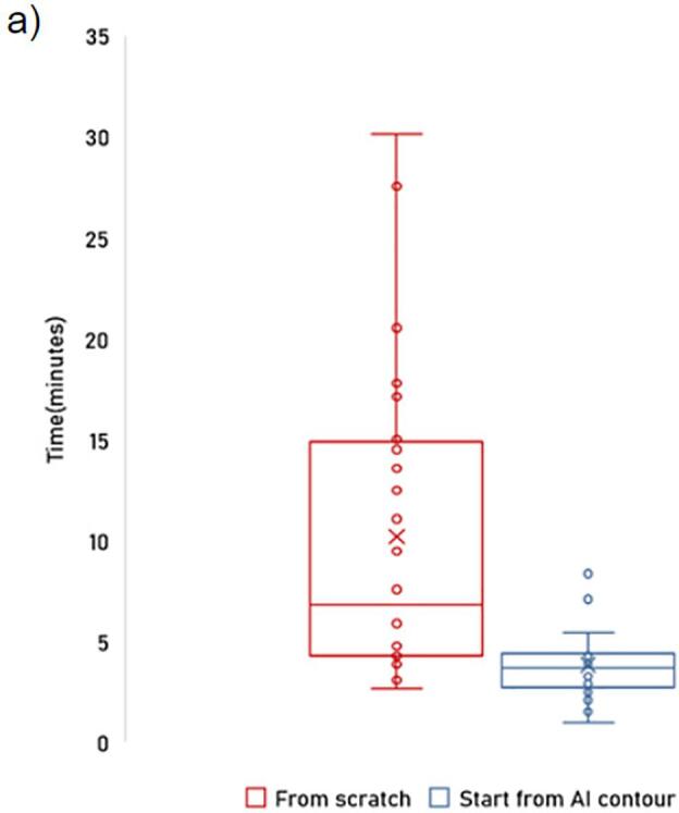
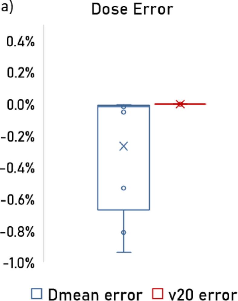
Test dataset metrics were DSC 61.71 ± 7.7 %, ASD 2.5 ± .87 mm, and HD95 7.0 ± 2.3 mm. AI segmented contours showed dosimetric similarity to expert physician's contours. Observer study indicated higher scores for AI contours (mean = 3.7) compared to inexperienced physicians' contours (mean = 3.1). Inexperienced physicians improved scores to 3.7 when starting with AI contours.
The proposed model achieved good quality IPA contours to improve uniformity of segmentation and to facilitate introduction of standardized IPA segmentation into clinical trials and practice.
放射性勃起功能障碍(RiED)通常会影响前列腺癌患者,促使各机构开展临床试验,探索对内阴部动脉(IPA)进行剂量 sparing 以保留性功能。IPA 分割具有挑战性,传统上不被视为危及器官(OAR)。本研究提出了一种用于 IPA 的深度学习(DL)自动分割模型,使用计算机断层扫描(CT)和磁共振成像(MRI)或仅使用 CT,以适应不同的临床实践。
本研究共招募了 86 例有 CT 和 MRI 图像以及有噪声的 IPA 标签的患者。我们将数据分别分为 42/14/30 用于模型训练、测试和临床观察者研究。该模型有三项主要创新:1)我们设计了一种带有挤压与激励块和模态注意力的架构,用于有效特征提取和生成准确分割,2)使用了一种新颖的损失函数,用于在有噪声标签的情况下有效训练模型,3)使用模态丢弃策略使模型能够在没有 MRI 的情况下进行分割。
测试数据集指标为 DSC 61.71 ± 7.7%,ASD 2.5 ± 0.87 毫米,HD95 7.0 ± 2.3 毫米。人工智能分割的轮廓与专家医生的轮廓显示出剂量学相似性。观察者研究表明,与经验不足的医生的轮廓(平均值 = 3.1)相比,人工智能轮廓的得分更高(平均值 = 3.7)。经验不足的医生从人工智能轮廓开始时,得分提高到了 3.7。
所提出的模型实现了高质量的 IPA 轮廓,以提高分割的均匀性,并便于将标准化的 IPA 分割引入临床试验和实践。