• 文献检索
  • 文档翻译
  • 深度研究
  • 学术资讯
  • Suppr Zotero 插件Zotero 插件
  • 邀请有礼
  • 套餐&价格
  • 历史记录
应用&插件
Suppr Zotero 插件Zotero 插件浏览器插件Mac 客户端Windows 客户端微信小程序
定价
高级版会员购买积分包购买API积分包
服务
文献检索文档翻译深度研究API 文档MCP 服务
关于我们
关于 Suppr公司介绍联系我们用户协议隐私条款
关注我们

Suppr 超能文献

核心技术专利:CN118964589B侵权必究
粤ICP备2023148730 号-1Suppr @ 2026

文献检索

告别复杂PubMed语法,用中文像聊天一样搜索,搜遍4000万医学文献。AI智能推荐,让科研检索更轻松。

立即免费搜索

文件翻译

保留排版,准确专业,支持PDF/Word/PPT等文件格式,支持 12+语言互译。

免费翻译文档

深度研究

AI帮你快速写综述,25分钟生成高质量综述,智能提取关键信息,辅助科研写作。

立即免费体验

无中心体纺锤体在非洲爪蟾卵提取物中,沿染色体附近分支的微管成核组装。

Acentrosomal spindles assemble from branching microtubule nucleation near chromosomes in Xenopus laevis egg extract.

机构信息

Department of Chemical and Biological Engineering, Princeton University, Princeton, NJ, 08544, USA.

Lewis-Sigler Institute for Integrative Genomics, Princeton University, Princeton, NJ, 08544, USA.

出版信息

Nat Commun. 2023 Jun 21;14(1):3696. doi: 10.1038/s41467-023-39041-z.

DOI:10.1038/s41467-023-39041-z
PMID:37344488
原文链接:https://pmc.ncbi.nlm.nih.gov/articles/PMC10284841/
Abstract

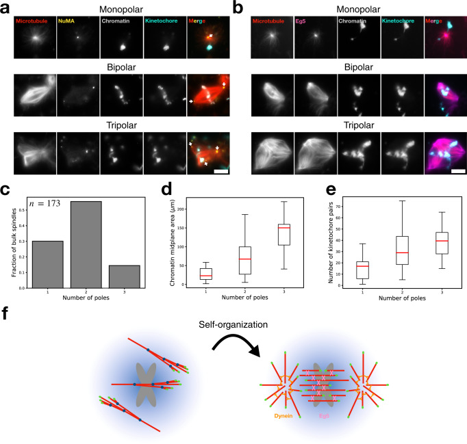
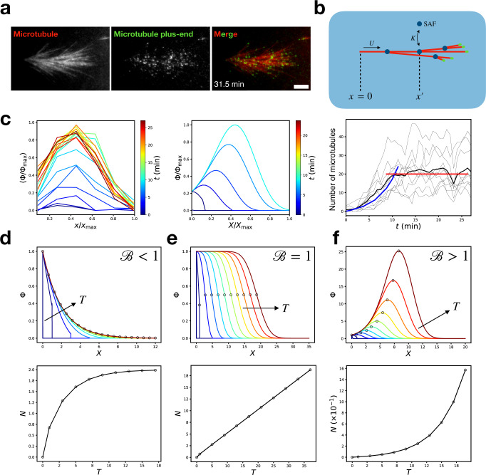
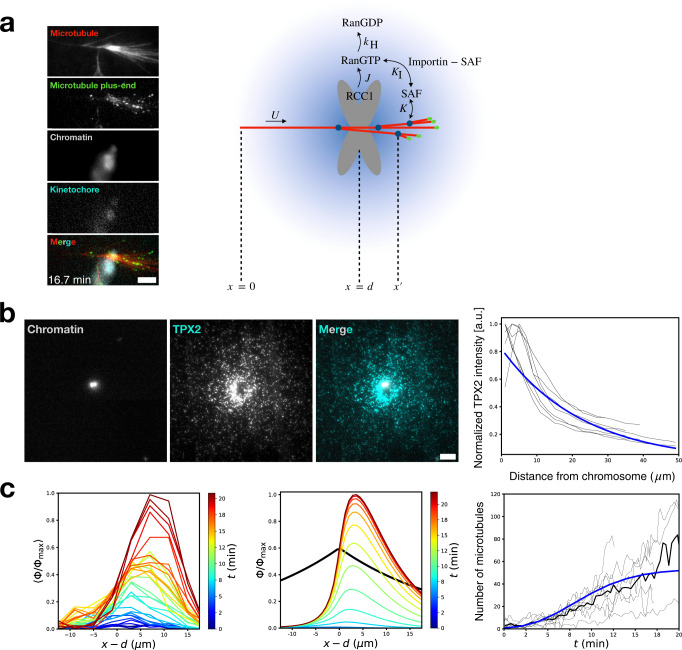
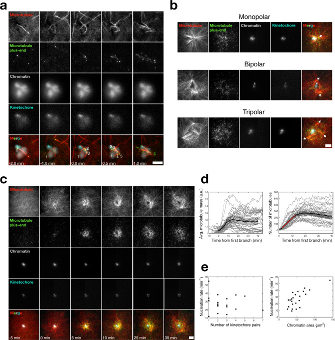
Microtubules are generated at centrosomes, chromosomes, and within spindles during cell division. Whereas microtubule nucleation at the centrosome is well characterized, much remains unknown about where, when, and how microtubules are nucleated at chromosomes. To address these questions, we reconstitute microtubule nucleation from purified chromosomes in meiotic Xenopus egg extract and find that chromosomes alone can form spindles. We visualize microtubule nucleation near chromosomes using total internal reflection fluorescence microscopy to find that this occurs through branching microtubule nucleation. By inhibiting molecular motors, we find that the organization of the resultant polar branched networks is consistent with a theoretical model where the effectors for branching nucleation are released by chromosomes, forming a concentration gradient that spatially biases branching microtbule nucleation. In the presence of motors, these branched networks are ultimately organized into functional spindles, where the number of emergent spindle poles scales with the number of chromosomes and total chromatin area.

摘要

微管在细胞分裂过程中在中心体、染色体和纺锤体中生成。虽然中心体处的微管核化已经得到很好的描述,但对于染色体处的微管核化在何处、何时以及如何发生,仍有很多未知。为了解决这些问题,我们在减数分裂的爪蟾卵提取物中从纯化的染色体中重新组装微管核化,发现仅染色体本身就可以形成纺锤体。我们使用全内反射荧光显微镜来观察染色体附近的微管核化,发现这是通过分支微管核化发生的。通过抑制分子马达,我们发现所得的极性分支网络的组织与分支核化的效应物由染色体释放的理论模型一致,形成了空间偏向分支微管核化的浓度梯度。在有马达的情况下,这些分支网络最终被组织成功能性的纺锤体,其中出现的纺锤体极的数量与染色体的数量和总染色质面积成比例。

https://cdn.ncbi.nlm.nih.gov/pmc/blobs/2e9e/10284841/4e6ce46395f6/41467_2023_39041_Fig6_HTML.jpg
https://cdn.ncbi.nlm.nih.gov/pmc/blobs/2e9e/10284841/a965b2c96a92/41467_2023_39041_Fig1_HTML.jpg
https://cdn.ncbi.nlm.nih.gov/pmc/blobs/2e9e/10284841/92ee6bff6bd5/41467_2023_39041_Fig2_HTML.jpg
https://cdn.ncbi.nlm.nih.gov/pmc/blobs/2e9e/10284841/3c7dd62b7eab/41467_2023_39041_Fig3_HTML.jpg
https://cdn.ncbi.nlm.nih.gov/pmc/blobs/2e9e/10284841/5adc83466765/41467_2023_39041_Fig4_HTML.jpg
https://cdn.ncbi.nlm.nih.gov/pmc/blobs/2e9e/10284841/0852f83993de/41467_2023_39041_Fig5_HTML.jpg
https://cdn.ncbi.nlm.nih.gov/pmc/blobs/2e9e/10284841/4e6ce46395f6/41467_2023_39041_Fig6_HTML.jpg

相似文献

1
Acentrosomal spindles assemble from branching microtubule nucleation near chromosomes in Xenopus laevis egg extract.无中心体纺锤体在非洲爪蟾卵提取物中,沿染色体附近分支的微管成核组装。
Nat Commun. 2023 Jun 21;14(1):3696. doi: 10.1038/s41467-023-39041-z.
2
Visualizing and Analyzing Branching Microtubule Nucleation Using Meiotic Xenopus Egg Extracts and TIRF Microscopy.利用非洲爪蟾卵母细胞减数分裂提取物和全内反射荧光显微镜观察及分析分支微管成核
Methods Mol Biol. 2016;1413:77-85. doi: 10.1007/978-1-4939-3542-0_6.
3
Multiple motors cooperate to establish and maintain acentrosomal spindle bipolarity in oocyte meiosis.多个马达协同作用以建立和维持卵母细胞减数分裂中的无中心体纺锤体双极性。
Elife. 2022 Feb 11;11:e72872. doi: 10.7554/eLife.72872.
4
Augmin promotes meiotic spindle formation and bipolarity in Xenopus egg extracts.Augmin 促进了非洲爪蟾卵提取物中减数分裂纺锤体的形成和两极体的形成。
Proc Natl Acad Sci U S A. 2011 Aug 30;108(35):14473-8. doi: 10.1073/pnas.1110412108. Epub 2011 Aug 15.
5
Autocatalytic microtubule nucleation determines the size and mass of egg extract spindles.自催化微管成核决定了卵提取物纺锤体的大小和质量。
Elife. 2018 Jan 11;7:e31149. doi: 10.7554/eLife.31149.
6
Spindle assembly in Xenopus egg extracts: respective roles of centrosomes and microtubule self-organization.非洲爪蟾卵提取物中的纺锤体组装:中心体与微管自组装的各自作用
J Cell Biol. 1997 Aug 11;138(3):615-28. doi: 10.1083/jcb.138.3.615.
7
Contribution of noncentrosomal microtubules to spindle assembly in Drosophila spermatocytes.非中心体微管对果蝇精母细胞纺锤体组装的贡献。
PLoS Biol. 2004 Jan;2(1):E8. doi: 10.1371/journal.pbio.0020008. Epub 2004 Jan 20.
8
Branching microtubule nucleation in Xenopus egg extracts mediated by augmin and TPX2.类星体卵提取物中由 augmin 和 TPX2 介导的分支微管核形成。
Cell. 2013 Feb 14;152(4):768-77. doi: 10.1016/j.cell.2012.12.044.
9
Making microtubules and mitotic spindles in cells without functional centrosomes.在没有功能性中心体的细胞中制造微管和有丝分裂纺锤体。
Curr Biol. 2006 Mar 21;16(6):564-9. doi: 10.1016/j.cub.2006.01.053.
10
Spatiotemporal organization of branched microtubule networks.分支微管网络的时空组织。
Elife. 2019 May 8;8:e43890. doi: 10.7554/eLife.43890.

引用本文的文献

1
HURP facilitates spindle assembly by stabilizing microtubules and working synergistically with TPX2.HURP 通过稳定微管并与 TPX2 协同作用来促进纺锤体的组装。
Nat Commun. 2024 Nov 8;15(1):9689. doi: 10.1038/s41467-024-53630-6.
2
Building on-chip cytoskeletal circuits via branched microtubule networks.通过分支微管网络构建片上细胞骨架电路。
Proc Natl Acad Sci U S A. 2024 Jan 23;121(4):e2315992121. doi: 10.1073/pnas.2315992121. Epub 2024 Jan 17.
3
HURP facilitates spindle assembly by stabilizing microtubules and working synergistically with TPX2.

本文引用的文献

1
Microtubule binding of the human augmin complex is directly controlled by importins and Ran-GTP.人类增敏复合物与微管的结合直接受输入蛋白和 Ran-GTP 的控制。
J Cell Sci. 2023 Jun 15;136(12). doi: 10.1242/jcs.261096. Epub 2023 Jun 26.
2
Microtubule nucleation from the fibrous corona by LIC1-pericentrin promotes chromosome congression.由 LIC1-中心体蛋白从纤维冠引发微管核形成促进染色体的向心移动。
Curr Biol. 2023 Mar 13;33(5):912-925.e6. doi: 10.1016/j.cub.2023.01.010. Epub 2023 Jan 30.
3
Morphological growth dynamics, mechanical stability, and active microtubule mechanics underlying spindle self-organization.
HURP通过稳定微管并与TPX2协同作用来促进纺锤体组装。
bioRxiv. 2023 Dec 18:2023.12.18.571906. doi: 10.1101/2023.12.18.571906.
4
Microtubule nucleation for spindle assembly: one molecule at a time.微管成核用于纺锤体组装:一次一个分子。
Trends Biochem Sci. 2023 Sep;48(9):761-775. doi: 10.1016/j.tibs.2023.06.004. Epub 2023 Jul 21.
5
Augmin is a Ran-regulated spindle assembly factor.Augmin 是一种 Ran 调控的纺锤体组装因子。
J Biol Chem. 2023 Jun;299(6):104736. doi: 10.1016/j.jbc.2023.104736. Epub 2023 Apr 21.
纺锤体自组织的形态生长动力学、力学稳定性和活性微管力学。
Proc Natl Acad Sci U S A. 2022 Nov;119(44):e2209053119. doi: 10.1073/pnas.2209053119. Epub 2022 Oct 25.
4
Cross-linker design determines microtubule network organization by opposing motors.交联剂设计通过与马达相反的作用来决定微管网络的组织。
Proc Natl Acad Sci U S A. 2022 Aug 16;119(33):e2206398119. doi: 10.1073/pnas.2206398119. Epub 2022 Aug 12.
5
Reconstitution and mechanistic dissection of the human microtubule branching machinery.重建和机制剖析人类微管分支机器。
J Cell Biol. 2022 Jul 4;221(7). doi: 10.1083/jcb.202109053. Epub 2022 May 23.
6
Phase separation of TPX2 enhances and spatially coordinates microtubule nucleation.TPX2 的相分离增强并空间协调微管成核。
Nat Commun. 2020 Jan 14;11(1):270. doi: 10.1038/s41467-019-14087-0.
7
In vitro reconstitution of branching microtubule nucleation.分支微管成核的体外重建
Elife. 2020 Jan 14;9:e49769. doi: 10.7554/eLife.49769.
8
Biochemical reconstitution of branching microtubule nucleation.分支微管成核的生化重建
Elife. 2020 Jan 14;9:e49797. doi: 10.7554/eLife.49797.
9
Augmin accumulation on long-lived microtubules drives amplification and kinetochore-directed growth.Augmin 聚集在长寿命微管上驱动扩增和动粒定向生长。
J Cell Biol. 2019 Jul 1;218(7):2150-2168. doi: 10.1083/jcb.201805044. Epub 2019 May 21.
10
Spatiotemporal organization of branched microtubule networks.分支微管网络的时空组织。
Elife. 2019 May 8;8:e43890. doi: 10.7554/eLife.43890.