Mycology, St John's Specialist Dermatology Laboratories, Synnovis, London, United Kingdom.
Br J Biomed Sci. 2023 Jun 7;80:11314. doi: 10.3389/bjbs.2023.11314. eCollection 2023.
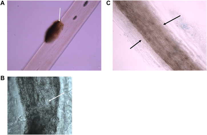
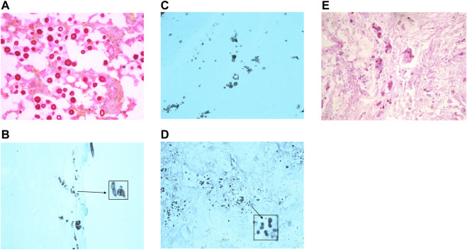
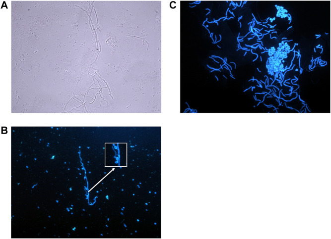
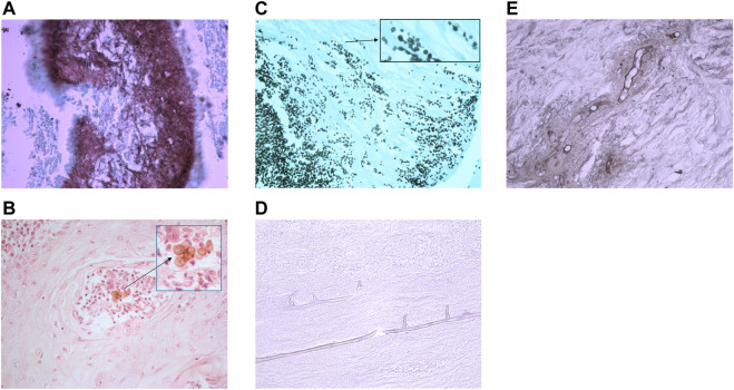
Diagnosis of superficial/cutaneous fungal infections from skin, hair and nail samples is generally achieved using microscopy and culture in a microbiology laboratory, however, any presentation that is unusual or subcutaneous is sampled by taking a biopsy. Using histological techniques a tissue biopsy enables a pathologist to perform a full examination of the skin structure, detect any inflammatory processes or the presence of an infectious agent or foreign body. Histopathological examination can give a presumptive diagnosis while a culture result is pending, and may provide valuable diagnostic information if culture fails. This review demonstrates how histopathology contributes to the diagnosis of fungal infections from the superficial to the life threatening.
从皮肤、毛发和指甲样本中诊断浅表/皮肤真菌感染通常在微生物实验室中使用显微镜和培养来完成,但是对于任何不常见或皮下的表现,都需要通过活检来取样。通过组织学技术,组织活检使病理学家能够对皮肤结构进行全面检查,检测任何炎症过程或感染因子或异物的存在。在等待培养结果时,组织病理学检查可以提供初步诊断,如果培养失败,它还可以提供有价值的诊断信息。这篇综述展示了组织病理学如何从浅表到危及生命的真菌感染的诊断中做出贡献。