• 文献检索
  • 文档翻译
  • 深度研究
  • 学术资讯
  • Suppr Zotero 插件Zotero 插件
  • 邀请有礼
  • 套餐&价格
  • 历史记录
应用&插件
Suppr Zotero 插件Zotero 插件浏览器插件Mac 客户端Windows 客户端微信小程序
定价
高级版会员购买积分包购买API积分包
服务
文献检索文档翻译深度研究API 文档MCP 服务
关于我们
关于 Suppr公司介绍联系我们用户协议隐私条款
关注我们

Suppr 超能文献

核心技术专利:CN118964589B侵权必究
粤ICP备2023148730 号-1Suppr @ 2026

文献检索

告别复杂PubMed语法,用中文像聊天一样搜索,搜遍4000万医学文献。AI智能推荐,让科研检索更轻松。

立即免费搜索

文件翻译

保留排版,准确专业,支持PDF/Word/PPT等文件格式,支持 12+语言互译。

免费翻译文档

深度研究

AI帮你快速写综述,25分钟生成高质量综述,智能提取关键信息,辅助科研写作。

立即免费体验

使用 b-张量编码和机器学习对白质组织病理学进行分化。

Differentiation of white matter histopathology using b-tensor encoding and machine learning.

机构信息

Instituto de Neurobiologia, Universidad Nacional Autónoma de Mexico, Querétaro, México.

Centro de Investigación en Matemáticas A.C., Guanajuato, México.

出版信息

PLoS One. 2023 Jun 23;18(6):e0282549. doi: 10.1371/journal.pone.0282549. eCollection 2023.

DOI:10.1371/journal.pone.0282549
PMID:37352195
原文链接:https://pmc.ncbi.nlm.nih.gov/articles/PMC10289327/
Abstract

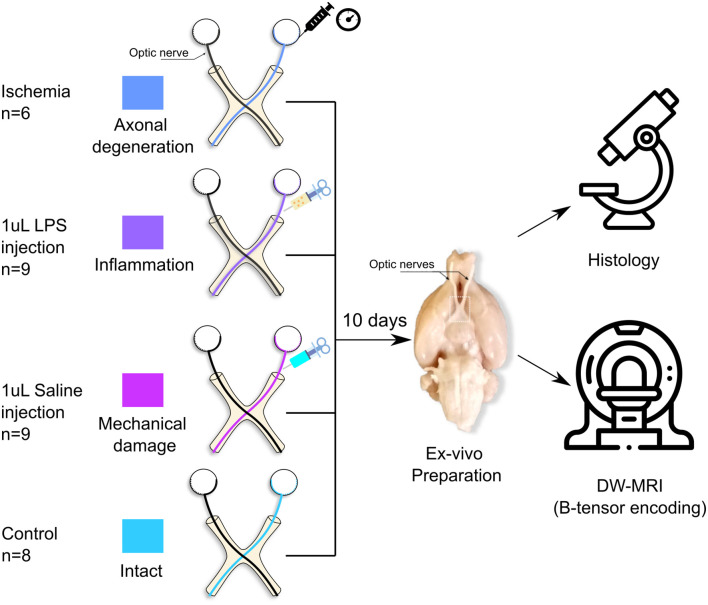
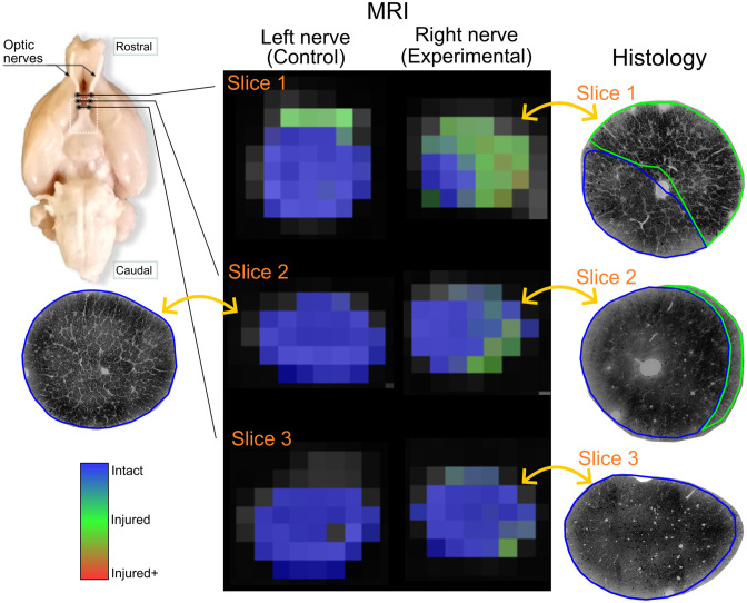
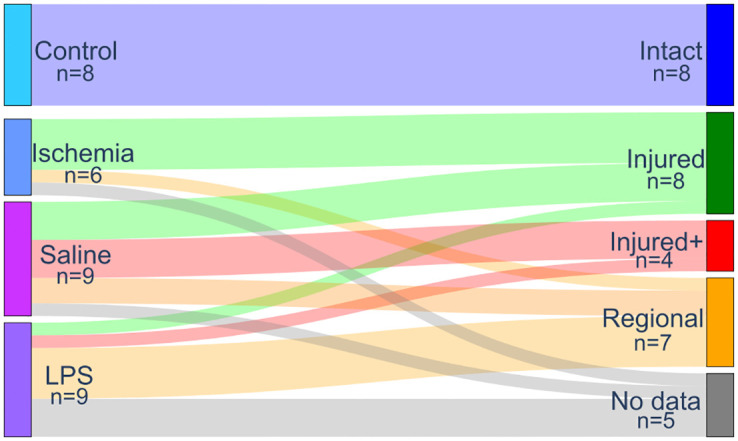
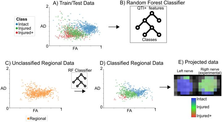
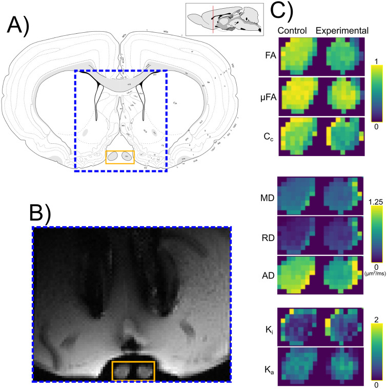
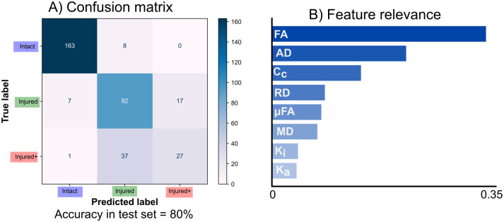
Diffusion-weighted magnetic resonance imaging (DW-MRI) is a non-invasive technique that is sensitive to microstructural geometry in neural tissue and is useful for the detection of neuropathology in research and clinical settings. Tensor-valued diffusion encoding schemes (b-tensor) have been developed to enrich the microstructural data that can be obtained through DW-MRI. These advanced methods have proven to be more specific to microstructural properties than conventional DW-MRI acquisitions. Additionally, machine learning methods are particularly useful for the study of multidimensional data sets. In this work, we have tested the reach of b-tensor encoding data analyses with machine learning in different histopathological scenarios. We achieved this in three steps: 1) We induced different levels of white matter damage in rodent optic nerves. 2) We obtained ex vivo DW-MRI data with b-tensor encoding schemes and calculated quantitative metrics using Q-space trajectory imaging. 3) We used a machine learning model to identify the main contributing features and built a voxel-wise probabilistic classification map of histological damage. Our results show that this model is sensitive to characteristics of microstructural damage. In conclusion, b-tensor encoded DW-MRI data analyzed with machine learning methods, have the potential to be further developed for the detection of histopathology and neurodegeneration.

摘要

扩散加权磁共振成像(DW-MRI)是一种非侵入性技术,对神经组织的微观结构几何形状敏感,可用于在研究和临床环境中检测神经病理学。张量值扩散编码方案(b-张量)已被开发出来,以丰富可以通过 DW-MRI 获得的微观结构数据。这些先进的方法已被证明比传统的 DW-MRI 采集更能反映微观结构特性。此外,机器学习方法在多维数据集的研究中特别有用。在这项工作中,我们已经测试了机器学习在不同组织病理学情况下对 b-张量编码数据分析的应用范围。我们分三个步骤实现了这一目标:1)我们在啮齿动物视神经中诱导不同程度的白质损伤。2)我们使用 b-张量编码方案获得离体 DW-MRI 数据,并使用 Q 空间轨迹成像计算定量指标。3)我们使用机器学习模型来识别主要贡献特征,并构建组织学损伤的体素级概率分类图。我们的结果表明,该模型对微观结构损伤的特征敏感。总之,用机器学习方法分析的 b-张量编码 DW-MRI 数据有可能进一步开发用于检测组织病理学和神经退行性变。

https://cdn.ncbi.nlm.nih.gov/pmc/blobs/6caa/10289327/98f82ffcd7ce/pone.0282549.g009.jpg
https://cdn.ncbi.nlm.nih.gov/pmc/blobs/6caa/10289327/78d8b3e29b44/pone.0282549.g001.jpg
https://cdn.ncbi.nlm.nih.gov/pmc/blobs/6caa/10289327/419a69120068/pone.0282549.g002.jpg
https://cdn.ncbi.nlm.nih.gov/pmc/blobs/6caa/10289327/a2bf7e1cc5ca/pone.0282549.g003.jpg
https://cdn.ncbi.nlm.nih.gov/pmc/blobs/6caa/10289327/2762519e5109/pone.0282549.g004.jpg
https://cdn.ncbi.nlm.nih.gov/pmc/blobs/6caa/10289327/01f013b1c076/pone.0282549.g005.jpg
https://cdn.ncbi.nlm.nih.gov/pmc/blobs/6caa/10289327/30cdb7aae167/pone.0282549.g006.jpg
https://cdn.ncbi.nlm.nih.gov/pmc/blobs/6caa/10289327/c14a2e4861e2/pone.0282549.g007.jpg
https://cdn.ncbi.nlm.nih.gov/pmc/blobs/6caa/10289327/77182de32556/pone.0282549.g008.jpg
https://cdn.ncbi.nlm.nih.gov/pmc/blobs/6caa/10289327/98f82ffcd7ce/pone.0282549.g009.jpg

相似文献

1
Differentiation of white matter histopathology using b-tensor encoding and machine learning.使用 b-张量编码和机器学习对白质组织病理学进行分化。
PLoS One. 2023 Jun 23;18(6):e0282549. doi: 10.1371/journal.pone.0282549. eCollection 2023.
2
Magic DIAMOND: Multi-fascicle diffusion compartment imaging with tensor distribution modeling and tensor-valued diffusion encoding.Magic DIAMOND:基于张量分布模型和张量值扩散编码的多束扩散隔室成像。
Med Image Anal. 2021 May;70:101988. doi: 10.1016/j.media.2021.101988. Epub 2021 Feb 4.
3
Q-space trajectory imaging for multidimensional diffusion MRI of the human brain.用于人类大脑多维扩散磁共振成像的Q空间轨迹成像
Neuroimage. 2016 Jul 15;135:345-62. doi: 10.1016/j.neuroimage.2016.02.039. Epub 2016 Feb 23.
4
Fiber estimation and tractography in diffusion MRI: development of simulated brain images and comparison of multi-fiber analysis methods at clinical b-values.扩散磁共振成像中的纤维估计与纤维束成像:模拟脑图像的开发及临床b值下多纤维分析方法的比较
Neuroimage. 2015 Apr 1;109:341-56. doi: 10.1016/j.neuroimage.2014.12.060. Epub 2014 Dec 30.
5
A comparison between diffusion tensor imaging and generalized q-sampling imaging in the age prediction of healthy adults via machine learning approaches.基于机器学习方法的弥散张量成像与广义 q 采样成像在健康成年人年龄预测中的比较。
J Neural Eng. 2022 Feb 1;19(1). doi: 10.1088/1741-2552/ac4bfe.
6
Texture analysis in brain T2 and diffusion MRI differentiates histology-verified grey and white matter pathology types in multiple sclerosis.脑 T2 和弥散 MRI 的纹理分析可区分多发性硬化症中经组织学证实的灰质和白质病变类型。
J Neurosci Methods. 2022 Sep 1;379:109671. doi: 10.1016/j.jneumeth.2022.109671. Epub 2022 Jul 9.
7
Deep learning reveals untapped information for local white-matter fiber reconstruction in diffusion-weighted MRI.深度学习揭示了弥散加权 MRI 中局部白质纤维重建的未被挖掘信息。
Magn Reson Imaging. 2019 Oct;62:220-227. doi: 10.1016/j.mri.2019.07.012. Epub 2019 Jul 16.
8
Diffusion tensor imaging analysis of rheumatoid arthritis patients with neuropsychiatric features to determine the alteration of white matter integrity due to vascular events.弥散张量成像分析伴神经精神症状的类风湿关节炎患者,以确定血管事件导致的白质完整性改变。
Clin Rheumatol. 2022 Oct;41(10):3169-3177. doi: 10.1007/s10067-022-06262-4. Epub 2022 Jun 25.
9
Machine-learning classification of 22q11.2 deletion syndrome: A diffusion tensor imaging study.22q11.2 缺失综合征的机器学习分类:一项弥散张量成像研究。
Neuroimage Clin. 2017 May 11;15:832-842. doi: 10.1016/j.nicl.2017.04.029. eCollection 2017.
10
Neural networks for parameter estimation in microstructural MRI: Application to a diffusion-relaxation model of white matter.神经网络在微观结构 MRI 参数估计中的应用:在白质扩散-弛豫模型中的应用。
Neuroimage. 2021 Dec 1;244:118601. doi: 10.1016/j.neuroimage.2021.118601. Epub 2021 Sep 22.

引用本文的文献

1
Spectral principal axis system (SPAS) and tuning of tensor-valued encoding for microscopic anisotropy and time-dependent diffusion in the rat brain.大鼠脑微观各向异性和时间依赖性扩散的光谱主轴系统(SPAS)及张量值编码的调谐
Imaging Neurosci (Camb). 2025 Jun 11;3. doi: 10.1162/IMAG.a.35. eCollection 2025.

本文引用的文献

1
Simultaneous superresolution reconstruction and distortion correction for single-shot EPI DWI using deep learning.使用深度学习对单次激发回波平面成像扩散加权成像进行同步超分辨率重建和畸变校正。
Magn Reson Med. 2023 Jun;89(6):2456-2470. doi: 10.1002/mrm.29601. Epub 2023 Jan 27.
2
Tissue optimization strategies for high-quality ex vivo diffusion imaging.组织优化策略可提高离体扩散成像的质量。
NMR Biomed. 2023 Mar;36(3):e4866. doi: 10.1002/nbm.4866. Epub 2022 Dec 4.
3
A review on investigation of the basic contrast mechanism underlying multidimensional diffusion MRI in assessment of neurological disorders.
关于在评估神经紊乱方面多维扩散 MRI 的基本对比机制的研究综述。
J Clin Neurosci. 2022 Aug;102:26-35. doi: 10.1016/j.jocn.2022.05.027. Epub 2022 Jun 10.
4
Mapping microglia and astrocyte activation in vivo using diffusion MRI.利用扩散磁共振成像在体内绘制小胶质细胞和星形胶质细胞的激活图谱。
Sci Adv. 2022 May 27;8(21):eabq2923. doi: 10.1126/sciadv.abq2923.
5
Reproducibility of the Standard Model of diffusion in white matter on clinical MRI systems.临床 MRI 系统中弥散的标准模型的可重复性。
Neuroimage. 2022 Aug 15;257:119290. doi: 10.1016/j.neuroimage.2022.119290. Epub 2022 May 8.
6
Myelin and non-myelin debris contribute to foamy macrophage formation after spinal cord injury.髓磷脂和非髓磷脂碎片在后脊髓损伤中导致泡沫状巨噬细胞的形成。
Neurobiol Dis. 2022 Feb;163:105608. doi: 10.1016/j.nbd.2021.105608. Epub 2021 Dec 31.
7
Correlation Tensor MRI deciphers underlying kurtosis sources in stroke.磁共振相关张量成像揭示中风中峰度源的基础信息。
Neuroimage. 2022 Feb 15;247:118833. doi: 10.1016/j.neuroimage.2021.118833. Epub 2021 Dec 18.
8
Histogram analysis of tensor-valued diffusion MRI in meningiomas: Relation to consistency, histological grade and type.张量值弥散 MRI 直方图分析在脑膜瘤中的应用:与一致性、组织学分级和类型的关系。
Neuroimage Clin. 2022;33:102912. doi: 10.1016/j.nicl.2021.102912. Epub 2021 Dec 13.
9
Neural networks for parameter estimation in microstructural MRI: Application to a diffusion-relaxation model of white matter.神经网络在微观结构 MRI 参数估计中的应用:在白质扩散-弛豫模型中的应用。
Neuroimage. 2021 Dec 1;244:118601. doi: 10.1016/j.neuroimage.2021.118601. Epub 2021 Sep 22.
10
On the potential for mapping apparent neural soma density via a clinically viable diffusion MRI protocol.通过一种临床可行的弥散磁共振成像协议来绘制明显的神经体素密度图的潜力。
Neuroimage. 2021 Oct 1;239:118303. doi: 10.1016/j.neuroimage.2021.118303. Epub 2021 Jun 23.