State Key Laboratory of Cell Differentiation and Regulation, and.
Henan International Joint Laboratory of Pulmonary Fibrosis, College of Life Science, Henan Normal University, Xinxiang, Henan, China.
Am J Respir Cell Mol Biol. 2023 Oct;69(4):456-469. doi: 10.1165/rcmb.2023-0080OC.
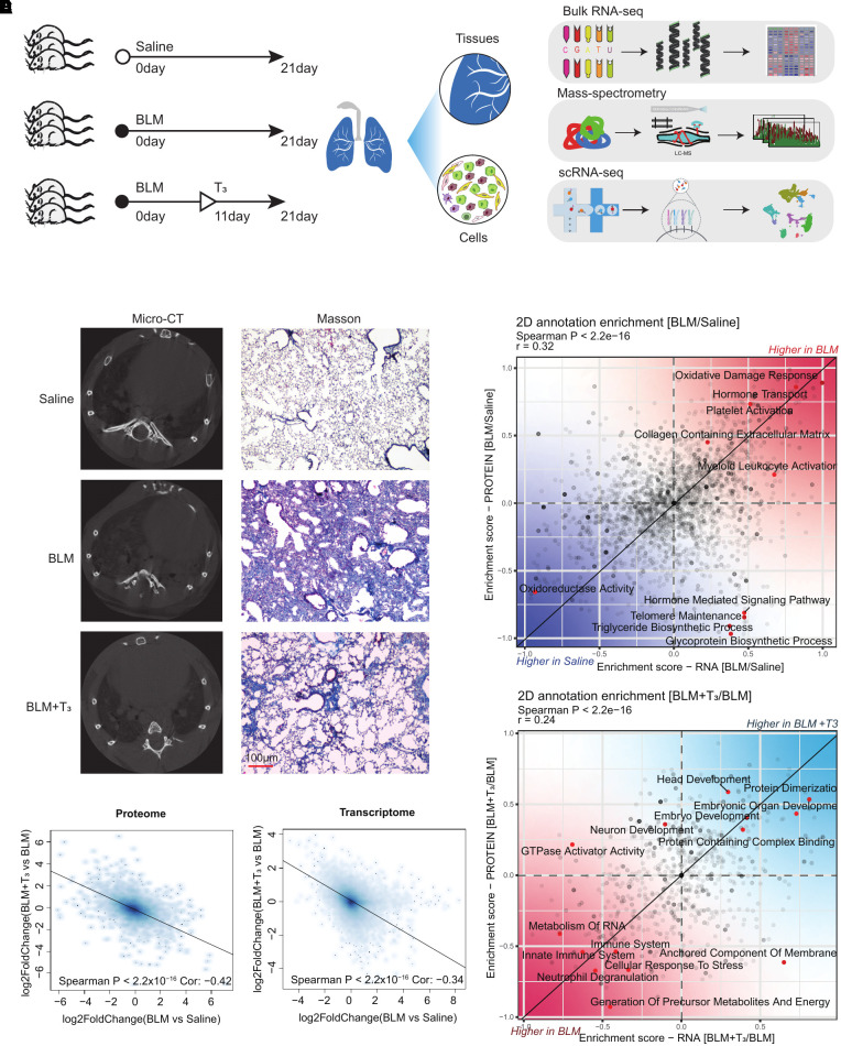
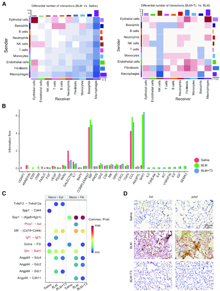
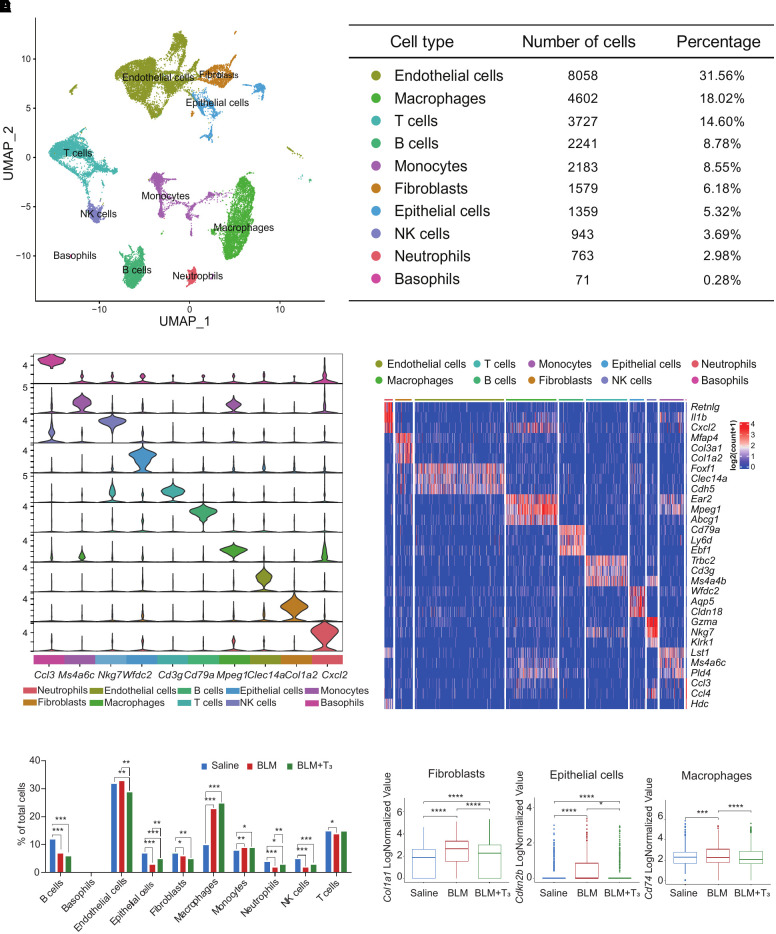
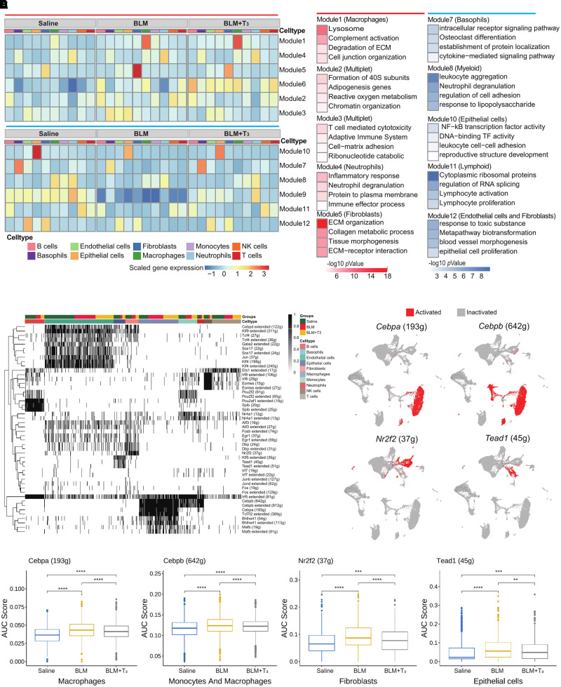
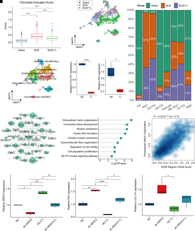
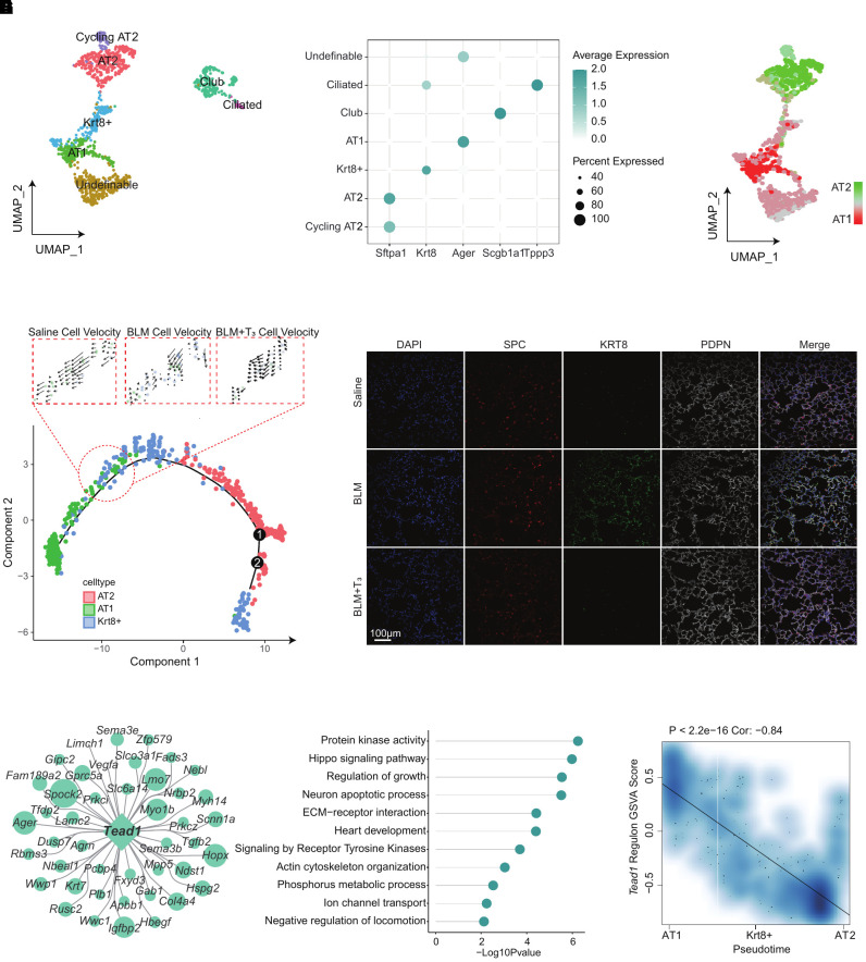
Idiopathic pulmonary fibrosis (IPF) is a progressive fatal interstitial lung disease without an effective cure. Herein, we explore the role of 3,5,3'-triiodothyronine (T) administration on lung alveolar regeneration and fibrosis at the single-cell level. T supplementation significantly altered the gene expression in fibrotic lung tissues. Immune cells were rapidly recruited into the lung after the injury; there were much more M2 macrophages than M1 macrophages in the lungs of bleomycin-treated mice; and M1 macrophages increased slightly, whereas M2 macrophages were significantly reduced after T treatment. T enhanced the resolution of pulmonary fibrosis by promoting the differentiation of transitional alveolar type II epithelial cells into alveolar type I epithelial cells and inhibiting fibroblast activation and extracellular matrix production potentially by regulation of . In addition, T regulated the crosstalk of macrophages with fibroblasts, and the signaling axis significantly facilitated the attenuation of fibrosis. The findings demonstrate that administration of a thyroid hormone promotes alveolar regeneration and resolves fibrosis mainly by regulation of the cellular state and cell-cell communication of alveolar epithelial cells, macrophages, and fibroblasts in mouse lungs in comprehensive ways.
特发性肺纤维化(IPF)是一种进行性致命的间质性肺疾病,目前尚无有效的治疗方法。在此,我们探讨了 3,5,3'-三碘甲状腺原氨酸(T)给药在肺泡再生和纤维化方面的作用,从单细胞水平上研究这一问题。T 补充显著改变了纤维化肺组织中的基因表达。损伤后,免疫细胞迅速被招募到肺部;博来霉素处理的小鼠肺部的 M2 巨噬细胞比 M1 巨噬细胞多得多;T 处理后,M1 巨噬细胞略有增加,而 M2 巨噬细胞明显减少。T 通过调节 促进过渡性肺泡 II 型上皮细胞向肺泡 I 型上皮细胞分化,抑制成纤维细胞激活和细胞外基质产生,从而增强肺纤维化的消退。此外,T 调节了巨噬细胞与成纤维细胞的串扰, 信号轴显著促进了纤维化的减弱。研究结果表明,甲状腺激素的给药通过调节细胞状态和细胞间通讯,以全面的方式促进肺泡再生和解决纤维化,主要涉及肺泡上皮细胞、巨噬细胞和成纤维细胞。