Kebiri Hamza, Gholipour Ali, Vasung Lana, Krsnik Željka, Karimi Davood, Cuadra Meritxell Bach
CIBM Center for Biomedical Imaging, Switzerland.
Department of Radiology, Lausanne University Hospital (CHUV) and University of Lausanne (UNIL), Lausanne, Switzerland.
bioRxiv. 2023 Jul 2:2023.07.01.547351. doi: 10.1101/2023.07.01.547351.
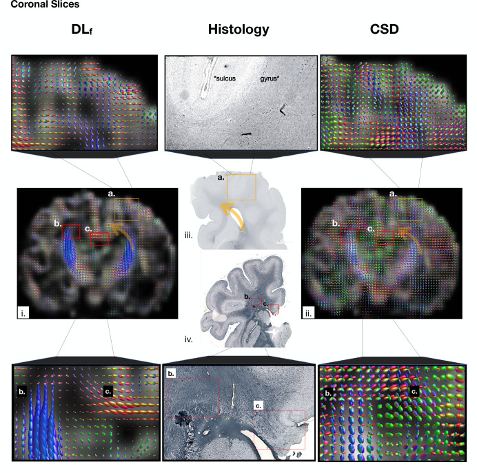
Diffusion-weighted magnetic resonance imaging (dMRI) is widely used to assess the brain white matter. Fiber orientation distribution functions (FODs) are a common way of representing the orientation and density of white matter fibers. However, with standard FOD computation methods, accurate estimation of FODs requires a large number of measurements that usually cannot be acquired for newborns and fetuses. We propose to overcome this limitation by using a deep learning method to map as few as six diffusion-weighted measurements to the target FOD. To train the model, we use the FODs computed using multi-shell high angular resolution measurements as target. Extensive quantitative evaluations show that the new deep learning method, using significantly fewer measurements, achieves comparable or superior results to standard methods such as Constrained Spherical Deconvolution. We demonstrate the generalizability of the new deep learning method across scanners, acquisition protocols, and anatomy on two clinical datasets of newborns and fetuses. Additionally, we compute agreement metrics within the HARDI newborn dataset, and validate fetal FODs with post-mortem histological data. The results of this study show the advantage of deep learning in inferring the microstructure of the developing brain from in-vivo dMRI measurements that are often very limited due to subject motion and limited acquisition times, but also highlight the intrinsic limitations of dMRI in the analysis of the developing brain microstructure. These findings, therefore, advocate for the need for improved methods that are tailored to studying the early development of human brain.
扩散加权磁共振成像(dMRI)被广泛用于评估脑白质。纤维方向分布函数(FODs)是表示白质纤维方向和密度的常用方法。然而,使用标准的FOD计算方法时,准确估计FODs需要大量测量,而新生儿和胎儿通常无法获得这么多测量数据。我们建议通过使用深度学习方法将少至六个扩散加权测量值映射到目标FOD来克服这一限制。为了训练模型,我们使用通过多壳高角分辨率测量计算得到的FODs作为目标。广泛的定量评估表明,这种新的深度学习方法使用的测量数据显著减少,却能取得与约束球形反卷积等标准方法相当或更优的结果。我们在新生儿和胎儿的两个临床数据集上展示了这种新的深度学习方法在不同扫描仪、采集协议和解剖结构上的通用性。此外,我们计算了HARDI新生儿数据集中的一致性指标,并用死后组织学数据验证了胎儿FODs。本研究结果显示了深度学习在从因受试者运动和采集时间有限而往往非常受限的活体dMRI测量中推断发育中大脑微观结构方面的优势,但也凸显了dMRI在分析发育中大脑微观结构时的内在局限性。因此,这些发现主张需要改进方法以专门研究人类大脑的早期发育。