Hamdi Mohammed, Senan Ebrahim Mohammed, Jadhav Mukti E, Olayah Fekry, Awaji Bakri, Alalayah Khaled M
Department of Computer Science, Faculty of Computer Science and Information System, Najran University, Najran 66462, Saudi Arabia.
Department of Artificial Intelligence, Faculty of Computer Science and Information Technology, Alrazi University, Sana'a, Yemen.
Diagnostics (Basel). 2023 Jul 4;13(13):2258. doi: 10.3390/diagnostics13132258.
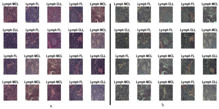
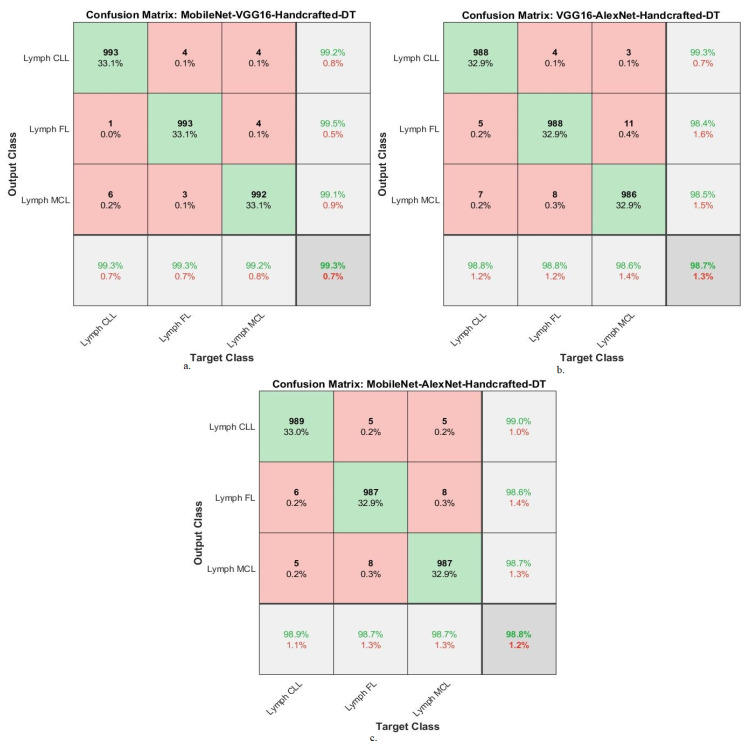
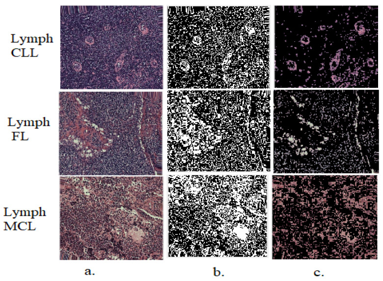
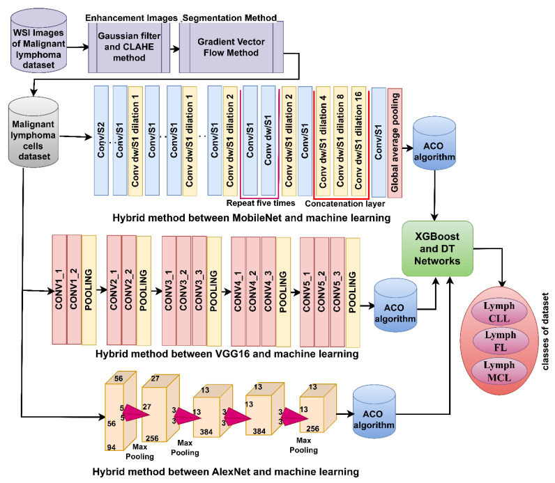
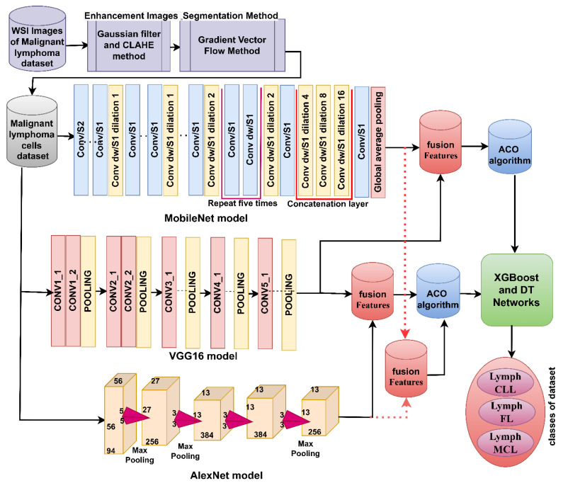
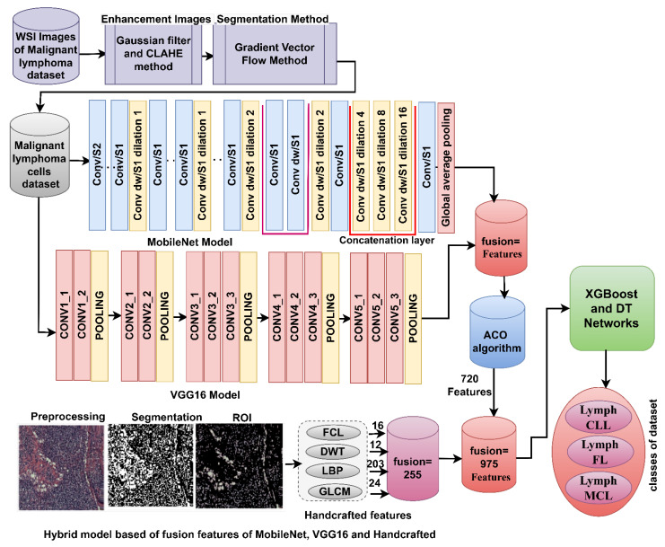
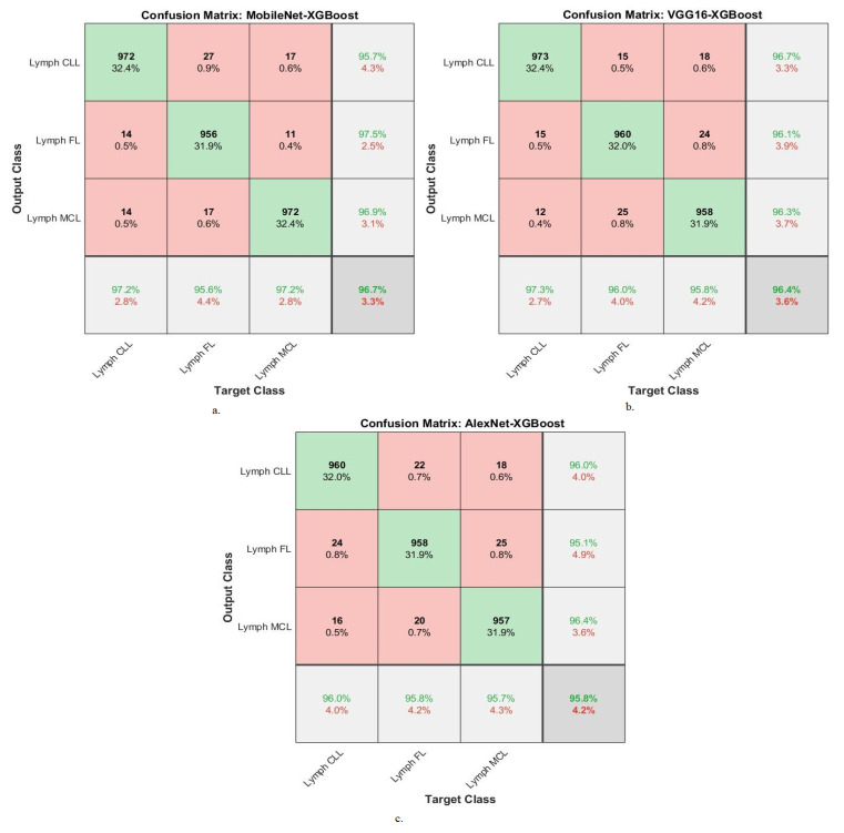
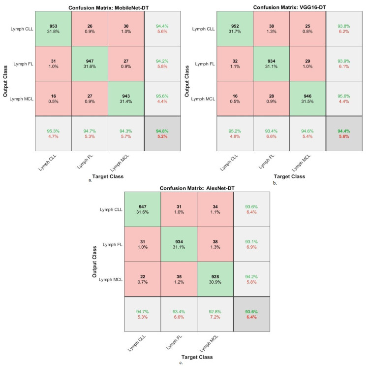
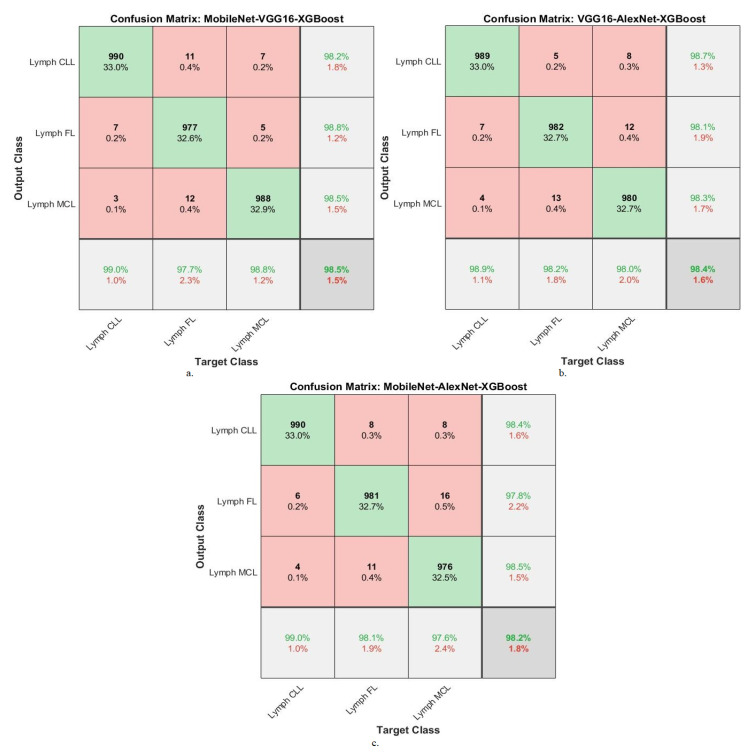
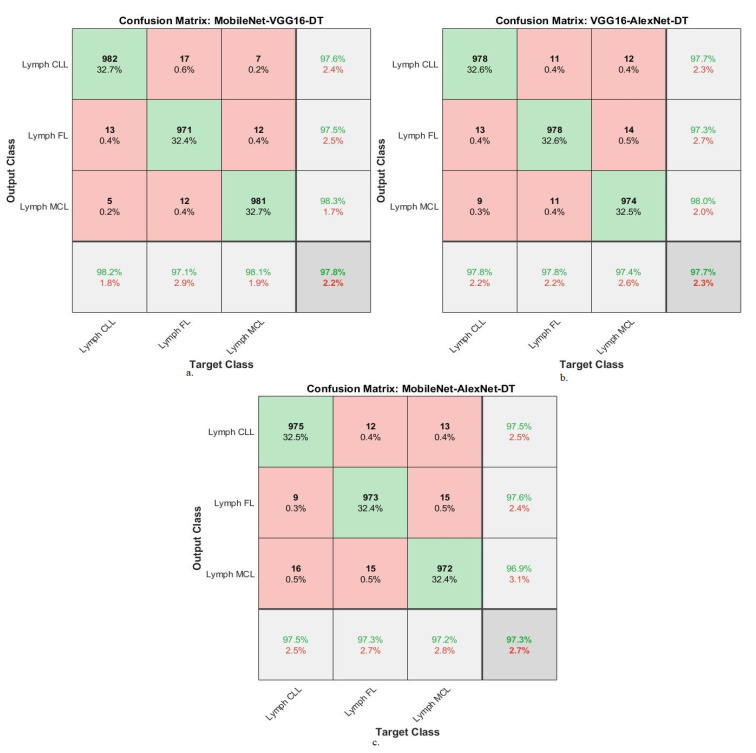
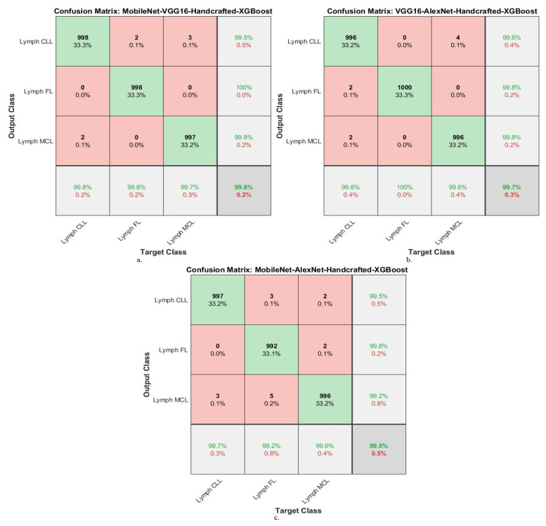
Malignant lymphoma is one of the most severe types of disease that leads to death as a result of exposure of lymphocytes to malignant tumors. The transformation of cells from indolent B-cell lymphoma to B-cell lymphoma (DBCL) is life-threatening. Biopsies taken from the patient are the gold standard for lymphoma analysis. Glass slides under a microscope are converted into whole slide images (WSI) to be analyzed by AI techniques through biomedical image processing. Because of the multiplicity of types of malignant lymphomas, manual diagnosis by pathologists is difficult, tedious, and subject to disagreement among physicians. The importance of artificial intelligence (AI) in the early diagnosis of malignant lymphoma is significant and has revolutionized the field of oncology. The use of AI in the early diagnosis of malignant lymphoma offers numerous benefits, including improved accuracy, faster diagnosis, and risk stratification. This study developed several strategies based on hybrid systems to analyze histopathological images of malignant lymphomas. For all proposed models, the images and extraction of malignant lymphocytes were optimized by the gradient vector flow (GVF) algorithm. The first strategy for diagnosing malignant lymphoma images relied on a hybrid system between three types of deep learning (DL) networks, XGBoost algorithms, and decision tree (DT) algorithms based on the GVF algorithm. The second strategy for diagnosing malignant lymphoma images was based on fusing the features of the MobileNet-VGG16, VGG16-AlexNet, and MobileNet-AlexNet models and classifying them by XGBoost and DT algorithms based on the ant colony optimization (ACO) algorithm. The color, shape, and texture features, which are called handcrafted features, were extracted by four traditional feature extraction algorithms. Because of the similarity in the biological characteristics of early-stage malignant lymphomas, the features of the fused MobileNet-VGG16, VGG16-AlexNet, and MobileNet-AlexNet models were combined with the handcrafted features and classified by the XGBoost and DT algorithms based on the ACO algorithm. We concluded that the performance of the two networks XGBoost and DT, with fused features between DL networks and handcrafted, achieved the best performance. The XGBoost network based on the fused features of MobileNet-VGG16 and handcrafted features resulted in an AUC of 99.43%, accuracy of 99.8%, precision of 99.77%, sensitivity of 99.7%, and specificity of 99.8%. This highlights the significant role of AI in the early diagnosis of malignant lymphoma, offering improved accuracy, expedited diagnosis, and enhanced risk stratification. This study highlights leveraging AI techniques and biomedical image processing; the analysis of whole slide images (WSI) converted from biopsies allows for improved accuracy, faster diagnosis, and risk stratification. The developed strategies based on hybrid systems, combining deep learning networks, XGBoost and decision tree algorithms, demonstrated promising results in diagnosing malignant lymphoma images. Furthermore, the fusion of handcrafted features with features extracted from DL networks enhanced the performance of the classification models.
恶性淋巴瘤是淋巴细胞遭受恶性肿瘤侵袭而导致死亡的最严重疾病类型之一。惰性B细胞淋巴瘤向弥漫性大B细胞淋巴瘤(DBCL)的细胞转化会危及生命。取自患者的活检样本是淋巴瘤分析的金标准。显微镜下的载玻片被转换为全切片图像(WSI),通过生物医学图像处理由人工智能技术进行分析。由于恶性淋巴瘤类型多样,病理学家进行人工诊断困难、繁琐,且医生之间容易出现分歧。人工智能(AI)在恶性淋巴瘤早期诊断中的重要性显著,它彻底改变了肿瘤学领域。在恶性淋巴瘤早期诊断中使用人工智能有诸多益处,包括提高准确性、加快诊断速度和进行风险分层。本研究基于混合系统开发了多种策略来分析恶性淋巴瘤的组织病理学图像。对于所有提出的模型,通过梯度向量流(GVF)算法对恶性淋巴细胞的图像和提取进行了优化。诊断恶性淋巴瘤图像的第一种策略依赖于基于GVF算法的三种深度学习(DL)网络、XGBoost算法和决策树(DT)算法之间的混合系统。诊断恶性淋巴瘤图像的第二种策略基于融合MobileNet-VGG16、VGG16-AlexNet和MobileNet-AlexNet模型的特征,并基于蚁群优化(ACO)算法通过XGBoost和DT算法进行分类。颜色、形状和纹理特征,即所谓的手工特征,通过四种传统特征提取算法提取。由于早期恶性淋巴瘤在生物学特征上具有相似性,融合后的MobileNet-VGG16、VGG16-AlexNet和MobileNet-AlexNet模型的特征与手工特征相结合,并基于ACO算法通过XGBoost和DT算法进行分类。我们得出结论,XGBoost和DT这两种网络在融合深度学习网络特征和手工特征后,取得了最佳性能。基于MobileNet-VGG融合特征和手工特征的XGBoost网络的曲线下面积(AUC)为99.43%,准确率为99.8%,精确率为99.77%,灵敏度为99.7%,特异性为99.8%。这突出了人工智能在恶性淋巴瘤早期诊断中的重要作用,可提高准确性、加快诊断速度并增强风险分层。本研究强调了利用人工智能技术和生物医学图像处理;对活检样本转换而来的全切片图像(WSI)进行分析可提高准确性、加快诊断速度并进行风险分层。基于混合系统开发的策略,结合深度学习网络、XGBoost和决策树算法,在诊断恶性淋巴瘤图像方面显示出了有前景的结果。此外,手工特征与从深度学习网络提取的特征相融合提高了分类模型的性能。