• 文献检索
  • 文档翻译
  • 深度研究
  • 学术资讯
  • Suppr Zotero 插件Zotero 插件
  • 邀请有礼
  • 套餐&价格
  • 历史记录
应用&插件
Suppr Zotero 插件Zotero 插件浏览器插件Mac 客户端Windows 客户端微信小程序
定价
高级版会员购买积分包购买API积分包
服务
文献检索文档翻译深度研究API 文档MCP 服务
关于我们
关于 Suppr公司介绍联系我们用户协议隐私条款
关注我们

Suppr 超能文献

核心技术专利:CN118964589B侵权必究
粤ICP备2023148730 号-1Suppr @ 2026

文献检索

告别复杂PubMed语法,用中文像聊天一样搜索,搜遍4000万医学文献。AI智能推荐,让科研检索更轻松。

立即免费搜索

文件翻译

保留排版,准确专业,支持PDF/Word/PPT等文件格式,支持 12+语言互译。

免费翻译文档

深度研究

AI帮你快速写综述,25分钟生成高质量综述,智能提取关键信息,辅助科研写作。

立即免费体验

基于混合特征的用于早期检测及区分肺炎和肺结核的X射线图像分析多技术

Multi-Techniques for Analyzing X-ray Images for Early Detection and Differentiation of Pneumonia and Tuberculosis Based on Hybrid Features.

作者信息

Ahmed Ibrahim Abdulrab, Senan Ebrahim Mohammed, Shatnawi Hamzeh Salameh Ahmad, Alkhraisha Ziad Mohammad, Al-Azzam Mamoun Mohammad Ali

机构信息

Computer Department, Applied College, Najran University, Najran 66462, Saudi Arabia.

Department of Artificial Intelligence, Faculty of Computer Science and Information Technology, Alrazi University, Sana'a, Yemen.

出版信息

Diagnostics (Basel). 2023 Feb 20;13(4):814. doi: 10.3390/diagnostics13040814.

DOI:10.3390/diagnostics13040814
PMID:36832302
原文链接:https://pmc.ncbi.nlm.nih.gov/articles/PMC9955018/
Abstract

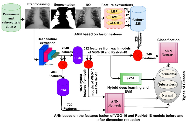
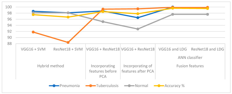
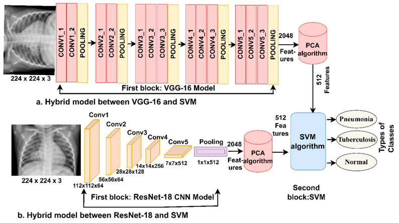
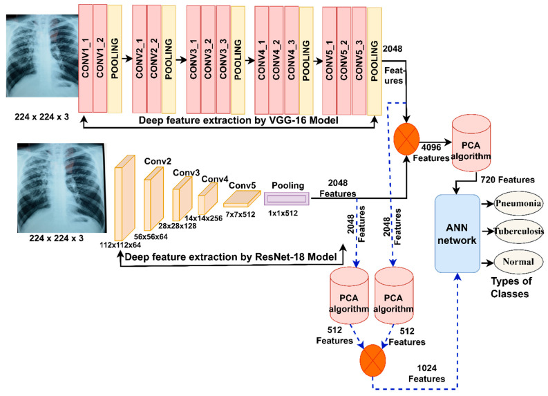
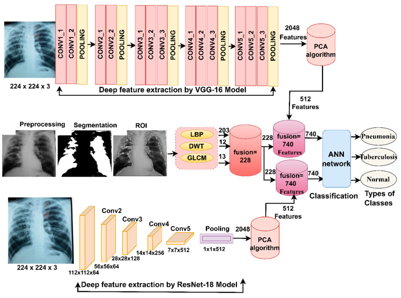
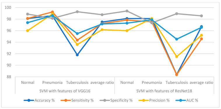
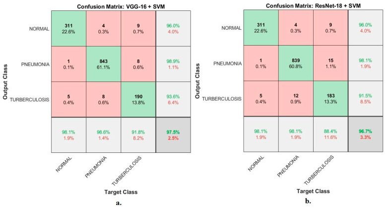
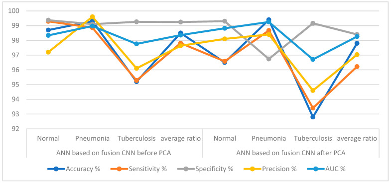
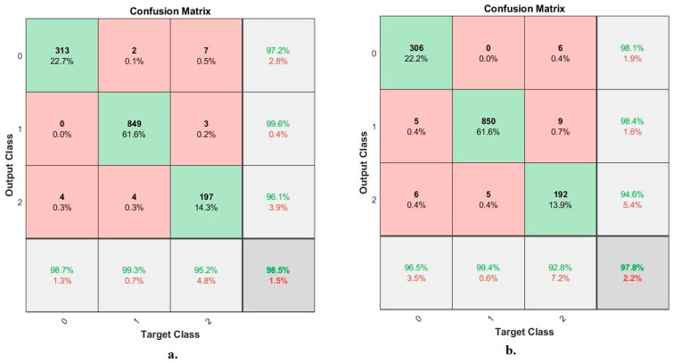
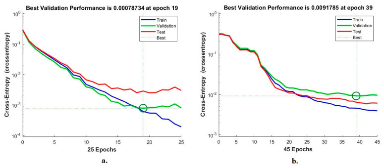
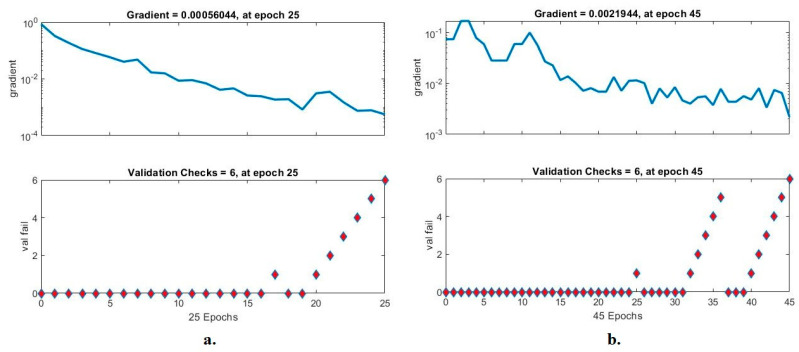
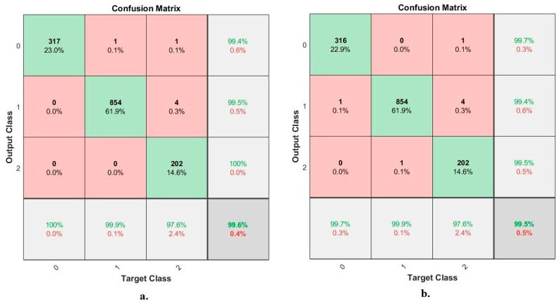
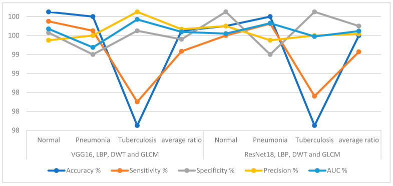
An infectious disease called tuberculosis (TB) exhibits pneumonia-like symptoms and traits. One of the most important methods for identifying and diagnosing pneumonia and tuberculosis is X-ray imaging. However, early discrimination is difficult for radiologists and doctors because of the similarities between pneumonia and tuberculosis. As a result, patients do not receive the proper care, which in turn does not prevent the disease from spreading. The goal of this study is to extract hybrid features using a variety of techniques in order to achieve promising results in differentiating between pneumonia and tuberculosis. In this study, several approaches for early identification and distinguishing tuberculosis from pneumonia were suggested. The first proposed system for differentiating between pneumonia and tuberculosis uses hybrid techniques, VGG16 + support vector machine (SVM) and ResNet18 + SVM. The second proposed system for distinguishing between pneumonia and tuberculosis uses an artificial neural network (ANN) based on integrating features of VGG16 and ResNet18, before and after reducing the high dimensions using the principal component analysis (PCA) method. The third proposed system for distinguishing between pneumonia and tuberculosis uses ANN based on integrating features of VGG16 and ResNet18 separately with handcrafted features extracted by local binary pattern (LBP), discrete wavelet transform (DWT) and gray level co-occurrence matrix (GLCM) algorithms. All the proposed systems have achieved superior results in the early differentiation between pneumonia and tuberculosis. An ANN based on the features of VGG16 with LBP, DWT and GLCM (LDG) reached an accuracy of 99.6%, sensitivity of 99.17%, specificity of 99.42%, precision of 99.63%, and an AUC of 99.58%.

摘要

一种名为肺结核(TB)的传染病表现出类似肺炎的症状和特征。X射线成像检查是鉴别诊断肺炎和肺结核最重要的手段之一。然而,由于肺炎和肺结核症状相似,放射科医生和临床医生很难进行早期鉴别。因此,患者无法得到恰当的治疗,疾病也无法得到有效控制。本研究旨在通过多种技术提取混合特征,以期在肺炎和肺结核的鉴别诊断中取得理想的结果。本研究提出了几种早期识别和区分肺结核与肺炎的方法。第一种用于区分肺炎和肺结核的系统采用混合技术,即VGG16+支持向量机(SVM)和ResNet18+SVM。第二种用于区分肺炎和肺结核的系统是基于VGG16和ResNet18特征融合的人工神经网络(ANN),并在使用主成分分析(PCA)方法进行高维降维前后进行特征融合。第三种用于区分肺炎和肺结核的系统是基于VGG16和ResNet18特征分别与通过局部二值模式(LBP)、离散小波变换(DWT)和灰度共生矩阵(GLCM)算法提取的手工特征进行融合的人工神经网络。所有提出的系统在肺炎和肺结核的早期鉴别中均取得了优异的结果。基于VGG16特征与LBP、DWT和GLCM(LDG)的人工神经网络的准确率达到99.6%,灵敏度达到99.17%,特异性达到99.42%,精确率达到99.63%,曲线下面积(AUC)达到99.58%。

https://cdn.ncbi.nlm.nih.gov/pmc/blobs/5a3d/9955018/511b731220f3/diagnostics-13-00814-g016.jpg
https://cdn.ncbi.nlm.nih.gov/pmc/blobs/5a3d/9955018/c630a42c74cc/diagnostics-13-00814-g001.jpg
https://cdn.ncbi.nlm.nih.gov/pmc/blobs/5a3d/9955018/4c5a45890e0f/diagnostics-13-00814-g002.jpg
https://cdn.ncbi.nlm.nih.gov/pmc/blobs/5a3d/9955018/df33e92658c9/diagnostics-13-00814-g003.jpg
https://cdn.ncbi.nlm.nih.gov/pmc/blobs/5a3d/9955018/b1c11eabe988/diagnostics-13-00814-g004.jpg
https://cdn.ncbi.nlm.nih.gov/pmc/blobs/5a3d/9955018/8e1e34bb5ef1/diagnostics-13-00814-g005.jpg
https://cdn.ncbi.nlm.nih.gov/pmc/blobs/5a3d/9955018/4c5ee6d9e3fd/diagnostics-13-00814-g006.jpg
https://cdn.ncbi.nlm.nih.gov/pmc/blobs/5a3d/9955018/a0f63e935af5/diagnostics-13-00814-g007.jpg
https://cdn.ncbi.nlm.nih.gov/pmc/blobs/5a3d/9955018/80896adf1957/diagnostics-13-00814-g008.jpg
https://cdn.ncbi.nlm.nih.gov/pmc/blobs/5a3d/9955018/b783a40b7cef/diagnostics-13-00814-g009.jpg
https://cdn.ncbi.nlm.nih.gov/pmc/blobs/5a3d/9955018/cce22b0139cc/diagnostics-13-00814-g010.jpg
https://cdn.ncbi.nlm.nih.gov/pmc/blobs/5a3d/9955018/29863e1d1d4c/diagnostics-13-00814-g011.jpg
https://cdn.ncbi.nlm.nih.gov/pmc/blobs/5a3d/9955018/af3f6df81cc7/diagnostics-13-00814-g012.jpg
https://cdn.ncbi.nlm.nih.gov/pmc/blobs/5a3d/9955018/dcac42c1daac/diagnostics-13-00814-g013.jpg
https://cdn.ncbi.nlm.nih.gov/pmc/blobs/5a3d/9955018/86e989bd9b38/diagnostics-13-00814-g014.jpg
https://cdn.ncbi.nlm.nih.gov/pmc/blobs/5a3d/9955018/1e96c0353d4e/diagnostics-13-00814-g015.jpg
https://cdn.ncbi.nlm.nih.gov/pmc/blobs/5a3d/9955018/511b731220f3/diagnostics-13-00814-g016.jpg

相似文献

1
Multi-Techniques for Analyzing X-ray Images for Early Detection and Differentiation of Pneumonia and Tuberculosis Based on Hybrid Features.基于混合特征的用于早期检测及区分肺炎和肺结核的X射线图像分析多技术
Diagnostics (Basel). 2023 Feb 20;13(4):814. doi: 10.3390/diagnostics13040814.
2
Analyzing Histological Images Using Hybrid Techniques for Early Detection of Multi-Class Breast Cancer Based on Fusion Features of CNN and Handcrafted.基于卷积神经网络(CNN)和手工特征融合的混合技术分析组织学图像以早期检测多类乳腺癌
Diagnostics (Basel). 2023 May 17;13(10):1753. doi: 10.3390/diagnostics13101753.
3
Early Diagnosis of Oral Squamous Cell Carcinoma Based on Histopathological Images Using Deep and Hybrid Learning Approaches.基于组织病理学图像,运用深度学习和混合学习方法对口腔鳞状细胞癌进行早期诊断。
Diagnostics (Basel). 2022 Aug 5;12(8):1899. doi: 10.3390/diagnostics12081899.
4
Hybrid and Deep Learning Approach for Early Diagnosis of Lower Gastrointestinal Diseases.混合与深度学习方法在胃肠道疾病早期诊断中的应用
Sensors (Basel). 2022 May 27;22(11):4079. doi: 10.3390/s22114079.
5
Automatic Analysis of MRI Images for Early Prediction of Alzheimer's Disease Stages Based on Hybrid Features of CNN and Handcrafted Features.基于卷积神经网络(CNN)混合特征和手工特征的MRI图像自动分析用于阿尔茨海默病阶段的早期预测
Diagnostics (Basel). 2023 May 8;13(9):1654. doi: 10.3390/diagnostics13091654.
6
Predicting of diabetic retinopathy development stages of fundus images using deep learning based on combined features.基于组合特征的深度学习预测眼底图像糖尿病视网膜病变的发展阶段。
PLoS One. 2023 Oct 20;18(10):e0289555. doi: 10.1371/journal.pone.0289555. eCollection 2023.
7
Hybrid Methods for Fundus Image Analysis for Diagnosis of Diabetic Retinopathy Development Stages Based on Fusion Features.基于融合特征的糖尿病视网膜病变发展阶段诊断的眼底图像分析混合方法
Diagnostics (Basel). 2023 Aug 28;13(17):2783. doi: 10.3390/diagnostics13172783.
8
Multi-Method Diagnosis of Blood Microscopic Sample for Early Detection of Acute Lymphoblastic Leukemia Based on Deep Learning and Hybrid Techniques.基于深度学习和混合技术的血液显微镜样本的多方法诊断,用于急性淋巴细胞白血病的早期检测。
Sensors (Basel). 2022 Feb 19;22(4):1629. doi: 10.3390/s22041629.
9
Multi-Method Analysis of Histopathological Image for Early Diagnosis of Oral Squamous Cell Carcinoma Using Deep Learning and Hybrid Techniques.基于深度学习和混合技术的口腔鳞状细胞癌早期诊断组织病理学图像多方法分析
Cancers (Basel). 2023 Oct 31;15(21):5247. doi: 10.3390/cancers15215247.
10
Hybrid Models Based on Fusion Features of a CNN and Handcrafted Features for Accurate Histopathological Image Analysis for Diagnosing Malignant Lymphomas.基于卷积神经网络融合特征与手工特征的混合模型用于准确的组织病理学图像分析以诊断恶性淋巴瘤
Diagnostics (Basel). 2023 Jul 4;13(13):2258. doi: 10.3390/diagnostics13132258.

引用本文的文献

1
Explainable deep-neural-network supported scheme for tuberculosis detection from chest radiographs.用于从胸部X光片中检测肺结核的可解释深度神经网络支持方案。
BMC Med Imaging. 2024 Feb 5;24(1):32. doi: 10.1186/s12880-024-01202-x.
2
Analysis of WSI Images by Hybrid Systems with Fusion Features for Early Diagnosis of Cervical Cancer.基于融合特征的混合系统对全切片图像进行分析以实现宫颈癌的早期诊断
Diagnostics (Basel). 2023 Jul 31;13(15):2538. doi: 10.3390/diagnostics13152538.
3
Hybrid Models Based on Fusion Features of a CNN and Handcrafted Features for Accurate Histopathological Image Analysis for Diagnosing Malignant Lymphomas.

本文引用的文献

1
Automatic Detection of Tuberculosis Using VGG19 with Seagull-Algorithm.使用带有海鸥算法的VGG19自动检测肺结核
Life (Basel). 2022 Nov 11;12(11):1848. doi: 10.3390/life12111848.
2
Explanatory classification of CXR images into COVID-19, Pneumonia and Tuberculosis using deep learning and XAI.使用深度学习和可解释性人工智能技术对 CXR 图像进行 COVID-19、肺炎和结核病的分类解释。
Comput Biol Med. 2022 Nov;150:106156. doi: 10.1016/j.compbiomed.2022.106156. Epub 2022 Oct 3.
3
Paving the way to a new class of efficient and safe tuberculosis vaccines: The role of c-di-AMP in immunity and virulence.
基于卷积神经网络融合特征与手工特征的混合模型用于准确的组织病理学图像分析以诊断恶性淋巴瘤
Diagnostics (Basel). 2023 Jul 4;13(13):2258. doi: 10.3390/diagnostics13132258.
4
Blood Slide Image Analysis to Classify WBC Types for Prediction Haematology Based on a Hybrid Model of CNN and Handcrafted Features.基于卷积神经网络(CNN)和手工特征混合模型的血液涂片图像分析,用于对白细胞类型进行分类以预测血液学情况。
Diagnostics (Basel). 2023 May 29;13(11):1899. doi: 10.3390/diagnostics13111899.
5
Automatic Classification of Colour Fundus Images for Prediction Eye Disease Types Based on Hybrid Features.基于混合特征的眼底彩色图像自动分类以预测眼病类型
Diagnostics (Basel). 2023 May 11;13(10):1706. doi: 10.3390/diagnostics13101706.
6
Automatic Analysis of MRI Images for Early Prediction of Alzheimer's Disease Stages Based on Hybrid Features of CNN and Handcrafted Features.基于卷积神经网络(CNN)混合特征和手工特征的MRI图像自动分析用于阿尔茨海默病阶段的早期预测
Diagnostics (Basel). 2023 May 8;13(9):1654. doi: 10.3390/diagnostics13091654.
7
Hybrid Techniques for the Diagnosis of Acute Lymphoblastic Leukemia Based on Fusion of CNN Features.基于卷积神经网络特征融合的急性淋巴细胞白血病诊断混合技术
Diagnostics (Basel). 2023 Mar 8;13(6):1026. doi: 10.3390/diagnostics13061026.
为新型高效安全的结核病疫苗铺平道路:环二腺苷酸在免疫和毒力中的作用
Mol Ther Nucleic Acids. 2022 Sep 12;30:13-14. doi: 10.1016/j.omtn.2022.08.034. eCollection 2022 Dec 13.
4
An efficient deep learning-based framework for tuberculosis detection using chest X-ray images.一种基于深度学习的高效框架,用于使用胸部X光图像检测肺结核。
Tuberculosis (Edinb). 2022 Sep;136:102234. doi: 10.1016/j.tube.2022.102234. Epub 2022 Jul 19.
5
Multi-branch fusion auxiliary learning for the detection of pneumonia from chest X-ray images.多分支融合辅助学习在胸部 X 射线图像肺炎检测中的应用。
Comput Biol Med. 2022 Aug;147:105732. doi: 10.1016/j.compbiomed.2022.105732. Epub 2022 Jun 15.
6
Urine biomarkers of pulmonary tuberculosis.尿生物标志物与肺结核。
Expert Rev Respir Med. 2022 Jun;16(6):615-621. doi: 10.1080/17476348.2022.2090341. Epub 2022 Jun 21.
7
Hybrid and Deep Learning Approach for Early Diagnosis of Lower Gastrointestinal Diseases.混合与深度学习方法在胃肠道疾病早期诊断中的应用
Sensors (Basel). 2022 May 27;22(11):4079. doi: 10.3390/s22114079.
8
Early Diagnosis of Brain Tumour MRI Images Using Hybrid Techniques between Deep and Machine Learning.基于深度学习和机器学习的混合技术对脑瘤 MRI 图像的早期诊断
Comput Math Methods Med. 2022 May 18;2022:8330833. doi: 10.1155/2022/8330833. eCollection 2022.
9
Tuberculosis Disease Diagnosis Based on an Optimized Machine Learning Model.基于优化机器学习模型的结核病诊断。
J Healthc Eng. 2022 Mar 21;2022:8950243. doi: 10.1155/2022/8950243. eCollection 2022.
10
Comparison of radiologist versus natural language processing-based image annotations for deep learning system for tuberculosis screening on chest radiographs.比较放射科医生与基于自然语言处理的图像标注对胸部 X 光片结核病筛查深度学习系统的影响。
Clin Imaging. 2022 Jul;87:34-37. doi: 10.1016/j.clinimag.2022.04.009. Epub 2022 Apr 25.