Pérez-Davila Sara, Garrido-Gulías Natalia, González-Rodríguez Laura, López-Álvarez Miriam, Serra Julia, López-Periago José Eugenio, González Pío
CINTECX, Universidade de Vigo, Grupo de Novos Materiais, 36310 Vigo, Spain.
Galicia Sur Health Research Institute (IIS Galicia Sur), SERGAS-UVIGO, 36213 Vigo, Spain.
Polymers (Basel). 2023 Jun 28;15(13):2849. doi: 10.3390/polym15132849.
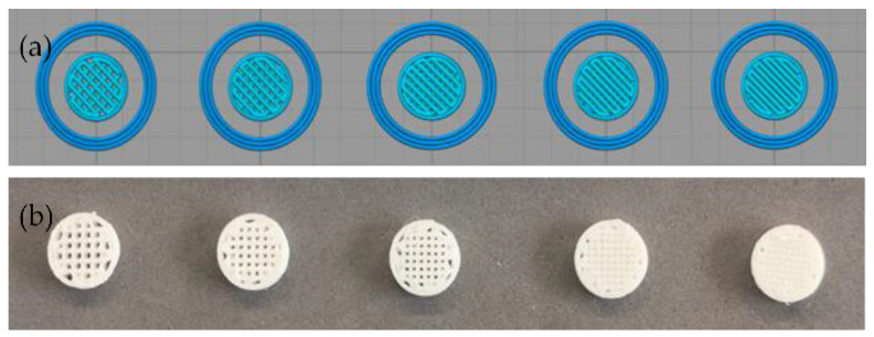
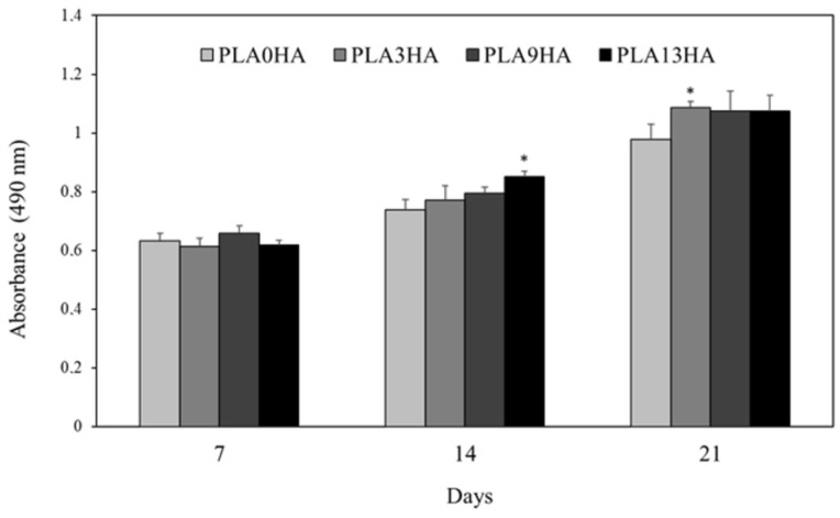
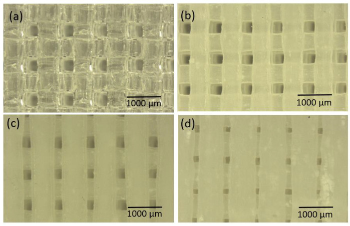
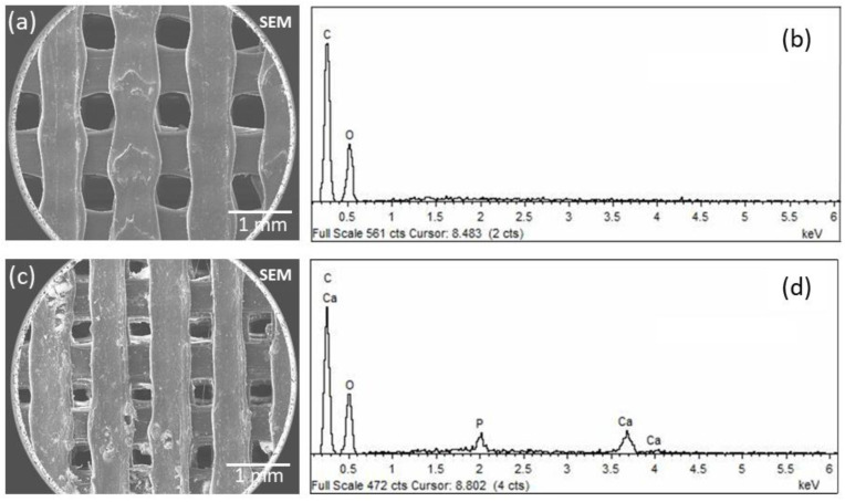
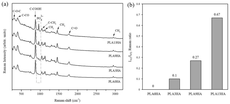
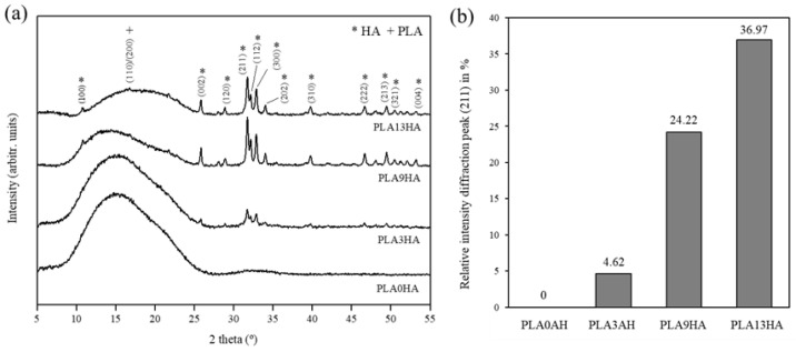
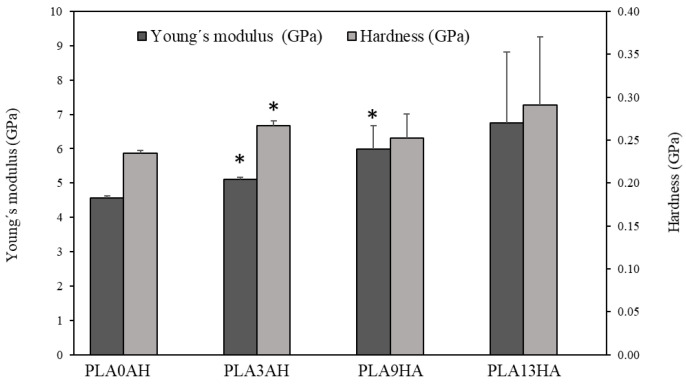
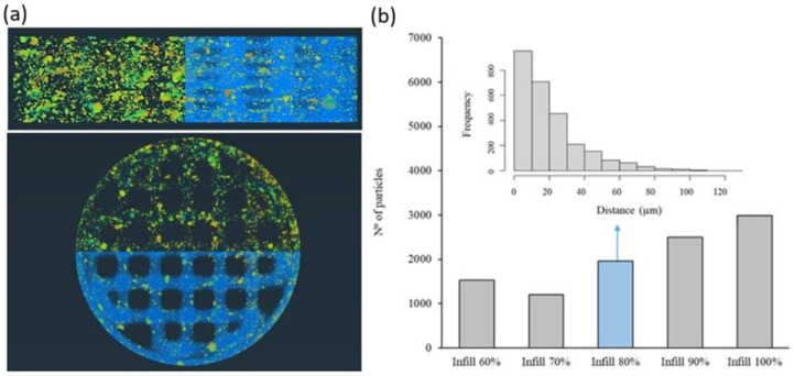
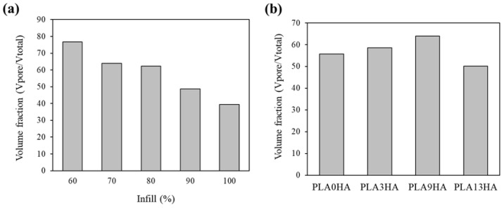
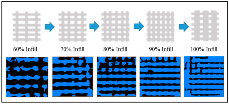
The reconstruction or regeneration of damaged bone tissue is one of the challenges of orthopedic surgery and tissue engineering. Among all strategies investigated, additive manufacturing by fused deposition modeling (3D-FDM printing) opens the possibility to obtain patient-specific scaffolds with controlled architectures. The present work evaluates in depth 3D direct printing, avoiding the need for a pre-fabricated filament, to obtain bone-related scaffolds from direct mixtures of polylactic acid (PLA) and hydroxyapatite (HA). For it, a systematic physicochemical characterization (SEM-EDS, FT-Raman, XRD, micro-CT and nanoindentation) was performed, using different PLA/HA ratios and percentages of infill. Results prove the versatility of this methodology with an efficient HA incorporation in the 3D-printed scaffolds up to 13 wt.% of the total mass and a uniform distribution of the HA particles in the scaffold at the macro level, both longitudinal and cross sections. Moreover, an exponential distribution of the HA particles from the surface toward the interior of the biocomposite cord (micro level), within the first 80 µm (10% of the entire cord diameter), is also confirmed, providing the scaffold with surface roughness and higher bioavailability. In relation to the pores, they can range in size from 250 to 850 µm and can represent a percentage, in relation to the total volume of the scaffold, from 24% up to 76%. The mechanical properties indicate an increase in Young's modulus with the HA content of up to ~50%, compared to the scaffolds without HA. Finally, the in vitro evaluation confirms MG63 cell proliferation on the 3D-printed PLA/HA scaffolds after up to 21 days of incubation.
受损骨组织的重建或再生是骨科手术和组织工程面临的挑战之一。在所有研究的策略中,熔融沉积建模增材制造(3D-FDM打印)为获得具有可控结构的患者特异性支架提供了可能性。本研究深入评估了3D直接打印,避免了对预制长丝的需求,以从聚乳酸(PLA)和羟基磷灰石(HA)的直接混合物中获得骨相关支架。为此,使用不同的PLA/HA比例和填充百分比进行了系统的物理化学表征(SEM-EDS、FT-拉曼、XRD、微型CT和纳米压痕)。结果证明了该方法的多功能性,在3D打印支架中HA的有效掺入量高达总质量的13 wt.%,并且在宏观水平上,在纵向和横截面上,HA颗粒在支架中均匀分布。此外,还证实了在生物复合线内部(微观水平),从表面到内部,HA颗粒在前80 µm(整个线直径的10%)内呈指数分布,为支架提供了表面粗糙度和更高的生物利用度。关于孔隙,其尺寸范围可以从250到850 µm,相对于支架的总体积,其占比可以从24%到76%。力学性能表明,与不含HA的支架相比,随着HA含量的增加,杨氏模量增加了约50%。最后,体外评估证实,在长达21天的孵育后,MG63细胞在3D打印的PLA/HA支架上增殖。