Neag Emily, Moceri Isabella, Harvey Faith, Udvadia Ava J, Bhattacharya Sanjoy K, Watson Fiona L
Bascom Palmer Eye Institute, Miller School of Medicine at University of Miami, Miami, FL, 33136, USA.
Miami Integrative Metabolomics Research Center, Miami, FL, 33136, USA.
Data Brief. 2023 Jun 14;49:109313. doi: 10.1016/j.dib.2023.109313. eCollection 2023 Aug.
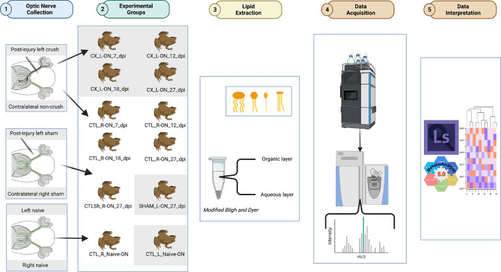
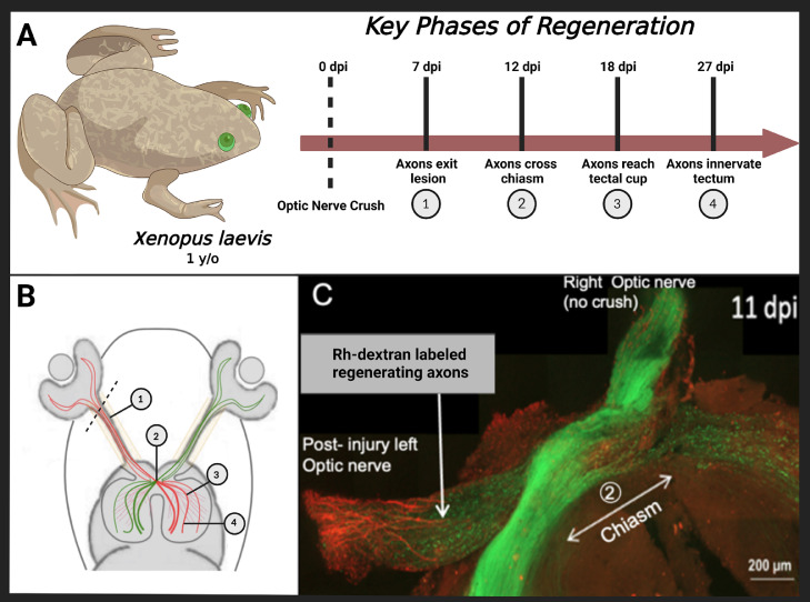
CNS injuries of the anuran amphibian, , are uniquely suited for studying the molecular compositions of neuronal regeneration of retinal ganglion cells (RGC) due to a functional recovery of optic axons disparate to adult mammalian analogues. RGCs and their optic nerve axons undergo irreversible neurodegeneration in glaucoma and associated optic neuropathies, resulting in blindness in mammals. Conversely, demonstrates RGC lifetime-spanning regenerative capabilities after optic nerve crush [1], inciting opportunities to compare de novo regeneration and develop efficient pharmaceutical approaches for vision restoration. Studies revealing lipidome alterations during optic nerve regeneration are sparse and could serve as a solid foundation for these underlying molecular changes. We profile the lipid changes in a transgenic line of 1 year old () frogs that were either left untreated (naïve) or had a monocular surgery of either a left optic crush injury (crush) or sham surgery (sham). Matching controls of uninjured right optic nerves were also collected (control). () frogs were allowed to recover for 7,12,18, and 27 days post optic nerve crush. Following euthanasia, the optic nerves were collected for lipidomic analysis. A modified Bligh and Dyer method [2] was used for lipid extraction, followed by untargeted mass spectrometry lipid profiling with a Q Exactive Orbitrap Mass Spectrometer coupled with a Vanquish Horizon Binary UHPLC LC-MS system (LC MS-MS). The raw scans were analyzed and quantified with LipidSearch 5.0 and the statistical analysis was conducted through Metaboanalyst 5.0. This data is available at Metabolomics Workbench, study ID [ST002414].
由于与成年哺乳动物类似物不同,无尾两栖动物的中枢神经系统损伤在视神经轴突功能恢复方面,特别适合用于研究视网膜神经节细胞(RGC)神经元再生的分子组成。在青光眼和相关视神经病变中,RGC及其视神经轴突会发生不可逆的神经变性,导致哺乳动物失明。相反,[研究对象]在视神经挤压后显示出RGC跨越生命周期的再生能力[1],这为比较从头再生和开发有效的视力恢复药物方法提供了机会。揭示视神经再生过程中脂质组变化的研究很少,可为这些潜在的分子变化提供坚实基础。我们分析了1岁[研究对象]转基因蛙系的脂质变化,这些蛙要么未接受治疗(未处理),要么接受了单眼手术,即左视神经挤压损伤(挤压)或假手术(假手术)。还收集了未受伤的右侧视神经的匹配对照(对照)。[研究对象]蛙在视神经挤压后分别恢复7、12、18和27天。安乐死后,收集视神经进行脂质组分析。采用改良的布利和戴尔方法[2]进行脂质提取,然后使用配备了Vanquish Horizon二元超高效液相色谱-质谱联用系统(LC MS-MS)的Q Exactive Orbitrap质谱仪进行非靶向质谱脂质谱分析。原始扫描数据用LipidSearch 5.0进行分析和定量,统计分析通过Metaboanalyst 5.0进行。该数据可在代谢组学工作台获取,研究ID为[ST002414]。