• 文献检索
  • 文档翻译
  • 深度研究
  • 学术资讯
  • Suppr Zotero 插件Zotero 插件
  • 邀请有礼
  • 套餐&价格
  • 历史记录
应用&插件
Suppr Zotero 插件Zotero 插件浏览器插件Mac 客户端Windows 客户端微信小程序
定价
高级版会员购买积分包购买API积分包
服务
文献检索文档翻译深度研究API 文档MCP 服务
关于我们
关于 Suppr公司介绍联系我们用户协议隐私条款
关注我们

Suppr 超能文献

核心技术专利:CN118964589B侵权必究
粤ICP备2023148730 号-1Suppr @ 2026

文献检索

告别复杂PubMed语法,用中文像聊天一样搜索,搜遍4000万医学文献。AI智能推荐,让科研检索更轻松。

立即免费搜索

文件翻译

保留排版,准确专业,支持PDF/Word/PPT等文件格式,支持 12+语言互译。

免费翻译文档

深度研究

AI帮你快速写综述,25分钟生成高质量综述,智能提取关键信息,辅助科研写作。

立即免费体验

无长突细胞中锌转运体3的选择性缺失可促进视网膜神经节细胞存活及损伤后视神经再生。

Selective deletion of zinc transporter 3 in amacrine cells promotes retinal ganglion cell survival and optic nerve regeneration after injury.

作者信息

Liu Zhe, Xue Jingfei, Liu Canying, Tang Jiahui, Wu Siting, Lin Jicheng, Han Jiaxu, Zhang Qi, Wu Caiqing, Huang Haishun, Zhao Ling, Zhuo Yehong, Li Yiqing

机构信息

State Key Laboratory of Ophthalmology, Zhongshan Ophthalmic Center, Sun Yat-sen University, Guangdong Provincial Key Laboratory of Ophthalmology and Visual Science, Guangzhou, Guangdong Province, China, Guangzhou.

出版信息

Neural Regen Res. 2023 Dec;18(12):2773-2780. doi: 10.4103/1673-5374.373660.

DOI:10.4103/1673-5374.373660
PMID:37449644
原文链接:https://pmc.ncbi.nlm.nih.gov/articles/PMC10358645/
Abstract

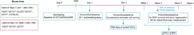
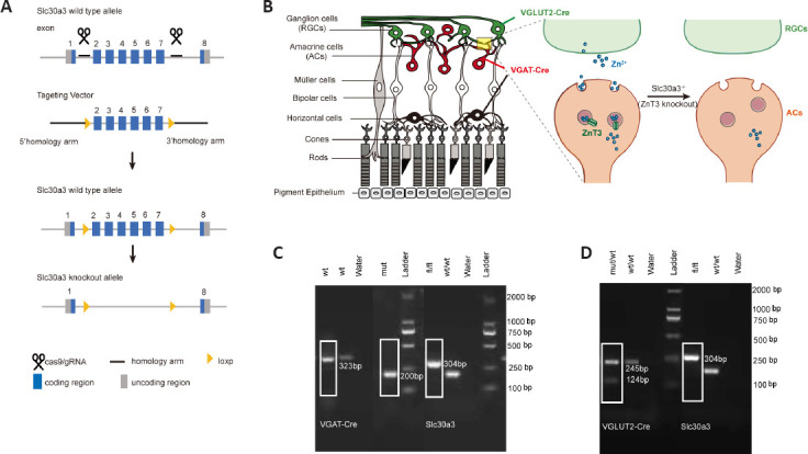
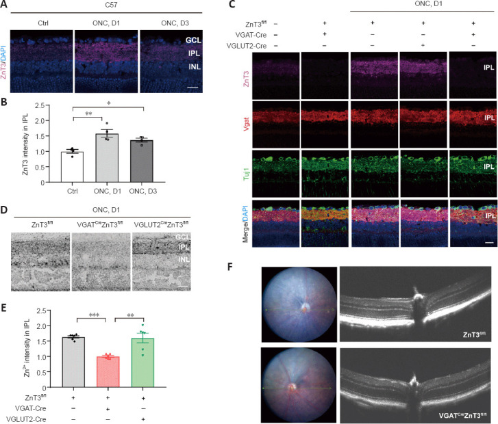
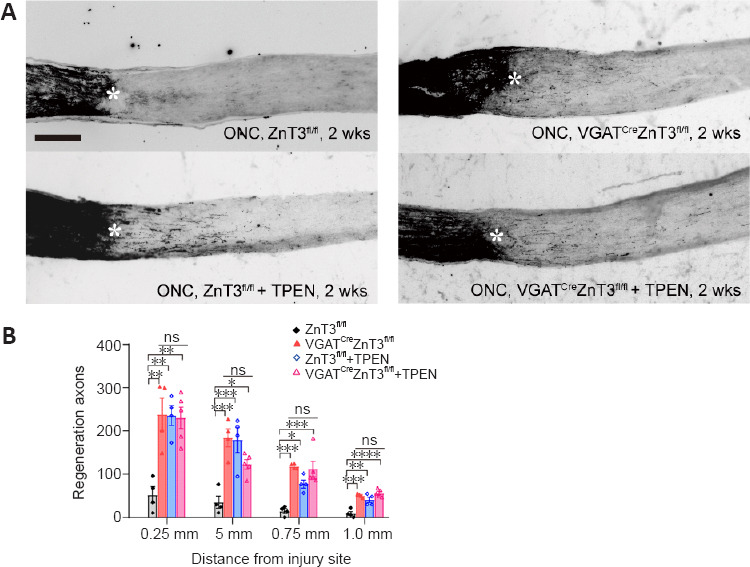
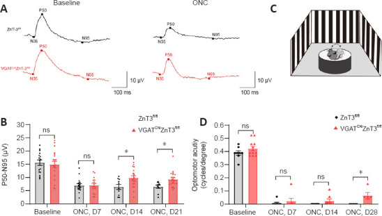
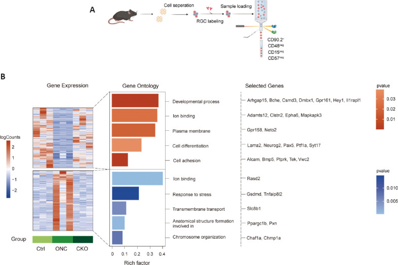
Vision depends on accurate signal conduction from the retina to the brain through the optic nerve, an important part of the central nervous system that consists of bundles of axons originating from retinal ganglion cells. The mammalian optic nerve, an important part of the central nervous system, cannot regenerate once it is injured, leading to permanent vision loss. To date, there is no clinical treatment that can regenerate the optic nerve and restore vision. Our previous study found that the mobile zinc (Zn) level increased rapidly after optic nerve injury in the retina, specifically in the vesicles of the inner plexiform layer. Furthermore, chelating Zn significantly promoted axonal regeneration with a long-term effect. In this study, we conditionally knocked out zinc transporter 3 (ZnT3) in amacrine cells or retinal ganglion cells to construct two transgenic mouse lines (VGATZnT3 and VGLUT2ZnT3, respectively). We obtained direct evidence that the rapidly increased mobile Zn in response to injury was from amacrine cells. We also found that selective deletion of ZnT3 in amacrine cells promoted retinal ganglion cell survival and axonal regeneration after optic nerve crush injury, improved retinal ganglion cell function, and promoted vision recovery. Sequencing analysis of reginal ganglion cells revealed that inhibiting the release of presynaptic Zn affected the transcription of key genes related to the survival of retinal ganglion cells in postsynaptic neurons, regulated the synaptic connection between amacrine cells and retinal ganglion cells, and affected the fate of retinal ganglion cells. These results suggest that amacrine cells release Zn to trigger transcriptomic changes related to neuronal growth and survival in reginal ganglion cells, thereby influencing the synaptic plasticity of retinal networks. These results make the theory of zinc-dependent retinal ganglion cell death more accurate and complete and provide new insights into the complex interactions between retinal cell networks.

摘要

视觉依赖于从视网膜通过视神经向大脑的准确信号传导,视神经是中枢神经系统的重要组成部分,由源自视网膜神经节细胞的轴突束组成。哺乳动物的视神经作为中枢神经系统的重要部分,一旦受损就无法再生,会导致永久性视力丧失。迄今为止,尚无能够使视神经再生并恢复视力的临床治疗方法。我们之前的研究发现,视网膜中视神经损伤后可移动锌(Zn)水平迅速升高,特别是在内网状层的囊泡中。此外,螯合锌可显著促进轴突再生且具有长期效果。在本研究中,我们有条件地敲除无长突细胞或视网膜神经节细胞中的锌转运体3(ZnT3),构建了两种转基因小鼠品系(分别为VGATZnT3和VGLUT2ZnT3)。我们获得了直接证据,表明损伤后迅速增加的可移动锌来自无长突细胞。我们还发现,选择性敲除无长突细胞中的ZnT3可促进视神经挤压伤后视网膜神经节细胞的存活和轴突再生,改善视网膜神经节细胞功能,并促进视力恢复。对视网膜神经节细胞的测序分析表明,抑制突触前锌的释放会影响突触后神经元中与视网膜神经节细胞存活相关的关键基因的转录,调节无长突细胞与视网膜神经节细胞之间的突触连接,并影响视网膜神经节细胞的命运。这些结果表明,无长突细胞释放锌以触发与视网膜神经节细胞中神经元生长和存活相关的转录组变化,从而影响视网膜网络的突触可塑性。这些结果使锌依赖性视网膜神经节细胞死亡理论更加准确和完整,并为视网膜细胞网络之间的复杂相互作用提供了新的见解。

https://cdn.ncbi.nlm.nih.gov/pmc/blobs/705e/10358645/c1c740bbef78/NRR-18-2773-g009.jpg
https://cdn.ncbi.nlm.nih.gov/pmc/blobs/705e/10358645/bdbbfa125f79/NRR-18-2773-g002.jpg
https://cdn.ncbi.nlm.nih.gov/pmc/blobs/705e/10358645/d31761e5ea54/NRR-18-2773-g003.jpg
https://cdn.ncbi.nlm.nih.gov/pmc/blobs/705e/10358645/87625313fb91/NRR-18-2773-g004.jpg
https://cdn.ncbi.nlm.nih.gov/pmc/blobs/705e/10358645/26710aff9af7/NRR-18-2773-g006.jpg
https://cdn.ncbi.nlm.nih.gov/pmc/blobs/705e/10358645/ae85dabd8d3e/NRR-18-2773-g007.jpg
https://cdn.ncbi.nlm.nih.gov/pmc/blobs/705e/10358645/204e2905c726/NRR-18-2773-g008.jpg
https://cdn.ncbi.nlm.nih.gov/pmc/blobs/705e/10358645/c1c740bbef78/NRR-18-2773-g009.jpg

相似文献

1
Selective deletion of zinc transporter 3 in amacrine cells promotes retinal ganglion cell survival and optic nerve regeneration after injury.无长突细胞中锌转运体3的选择性缺失可促进视网膜神经节细胞存活及损伤后视神经再生。
Neural Regen Res. 2023 Dec;18(12):2773-2780. doi: 10.4103/1673-5374.373660.
2
Mobile zinc increases rapidly in the retina after optic nerve injury and regulates ganglion cell survival and optic nerve regeneration.视神经损伤后,视网膜中的可移动锌迅速增加,并调节神经节细胞存活和视神经再生。
Proc Natl Acad Sci U S A. 2017 Jan 10;114(2):E209-E218. doi: 10.1073/pnas.1616811114. Epub 2017 Jan 3.
3
Reduced Zn promotes retinal ganglion cells survival and optic nerve regeneration after injury through inhibiting autophagy mediated by ROS/Nrf2.Zn 缺乏可通过抑制 ROS/Nrf2 介导的自噬促进损伤后视网膜神经节细胞的存活和视神经再生。
Free Radic Biol Med. 2024 Feb 20;212:415-432. doi: 10.1016/j.freeradbiomed.2023.12.008. Epub 2023 Dec 20.
4
Cell type-specific Nogo-A gene ablation promotes axonal regeneration in the injured adult optic nerve.细胞类型特异性的Nogo-A基因缺失促进成年受损视神经的轴突再生。
Cell Death Differ. 2015 Feb;22(2):323-35. doi: 10.1038/cdd.2014.147. Epub 2014 Sep 26.
5
Synaptic or Non-synaptic? Different Intercellular Interactions with Retinal Ganglion Cells in Optic Nerve Regeneration.突触还是非突触?视神经再生中与视网膜神经节细胞的不同细胞间相互作用。
Mol Neurobiol. 2022 May;59(5):3052-3072. doi: 10.1007/s12035-022-02781-y. Epub 2022 Mar 9.
6
Sphingosine 1-phosphate receptor 1 is required for retinal ganglion cell survival after optic nerve trauma.1-磷酸鞘氨醇受体1是视神经损伤后视网膜神经节细胞存活所必需的。
J Neurochem. 2016 Aug;138(4):571-86. doi: 10.1111/jnc.13701. Epub 2016 Jul 6.
7
Increased Mobile Zinc Regulates Retinal Ganglion Cell Survival via Activating Mitochondrial OMA1 and Integrated Stress Response.增加的可移动锌通过激活线粒体OMA1和综合应激反应来调节视网膜神经节细胞的存活。
Antioxidants (Basel). 2022 Oct 10;11(10):2001. doi: 10.3390/antiox11102001.
8
The basic science of optic nerve regeneration.视神经再生的基础科学。
Ann Transl Med. 2021 Aug;9(15):1276. doi: 10.21037/atm-20-5351.
9
GPR3 expression in retinal ganglion cells contributes to neuron survival and accelerates axonal regeneration after optic nerve crush in mice.GPR3 在视网膜神经节细胞中的表达有助于神经元存活,并加速小鼠视神经挤压后的轴突再生。
Neurobiol Dis. 2022 Oct 1;172:105811. doi: 10.1016/j.nbd.2022.105811. Epub 2022 Jul 7.
10
Microglial SIRT1 activation attenuates synapse loss in retinal inner plexiform layer via mTORC1 inhibition.小胶质细胞 SIRT1 的激活通过抑制 mTORC1 来减轻视网膜内丛状层的突触丢失。
J Neuroinflammation. 2023 Sep 5;20(1):202. doi: 10.1186/s12974-023-02886-8.

引用本文的文献

1
A role of pigment epithelium-derived factor in zinc-mediated mechanism of neurodegeneration in glaucoma.色素上皮衍生因子在青光眼锌介导的神经退行性变机制中的作用。
Commun Biol. 2025 Jul 1;8(1):965. doi: 10.1038/s42003-025-08370-8.
2
Inhibition of IGF-1Rα affects the differentiation fate of rat optic cup-derived retinal stem cells to retinal ganglion cells .抑制胰岛素样生长因子-1受体α(IGF-1Rα)会影响大鼠视杯来源的视网膜干细胞向视网膜神经节细胞的分化命运。
Int J Ophthalmol. 2025 Apr 18;18(4):582-589. doi: 10.18240/ijo.2025.04.03. eCollection 2025.
3
TPEN loaded poly (lactide-co-glycolide) nanoparticles promote neuroprotection and optic nerve regeneration.

本文引用的文献

1
Single-cell RNA sequencing reveals a landscape and targeted treatment of ferroptosis in retinal ischemia/reperfusion injury.单细胞 RNA 测序揭示了视网膜缺血/再灌注损伤中 ferroptosis 的全景和靶向治疗。
J Neuroinflammation. 2022 Oct 26;19(1):261. doi: 10.1186/s12974-022-02621-9.
2
Consequences of zinc deficiency on zinc localization, taurine transport, and zinc transporters in rat retina.锌缺乏对大鼠视网膜中锌定位、牛磺酸转运和锌转运体的影响。
Microsc Res Tech. 2022 Oct;85(10):3382-3390. doi: 10.1002/jemt.24193. Epub 2022 Jul 14.
3
-crosslinked hydrogel-induced experimental glaucoma model with persistent ocular hypertension and neurodegeneration.
负载三乙烯四胺的聚(丙交酯-乙交酯)纳米颗粒促进神经保护和视神经再生。
Mater Today Bio. 2025 Mar 14;32:101670. doi: 10.1016/j.mtbio.2025.101670. eCollection 2025 Jun.
4
Unlocking the potential for optic nerve regeneration over long distances: a multi-therapeutic intervention.释放视神经远距离再生的潜力:一种多疗法干预措施。
Front Neurol. 2025 Jan 9;15:1526973. doi: 10.3389/fneur.2024.1526973. eCollection 2024.
5
Modulating amacrine cell-derived dopamine signaling promotes optic nerve regeneration and preserves visual function.调节无长突细胞衍生的多巴胺信号促进视神经再生并维持视觉功能。
Sci Adv. 2024 Aug 2;10(31):eado0866. doi: 10.1126/sciadv.ado0866.
交联水凝胶诱导的持续性眼高压和神经退行性变实验性青光眼模型。
Biomater Sci. 2022 Aug 24;10(17):5006-5017. doi: 10.1039/d2bm00552b.
4
Core transcription programs controlling injury-induced neurodegeneration of retinal ganglion cells.核心转录程序控制视网膜神经节细胞损伤诱导的神经变性。
Neuron. 2022 Aug 17;110(16):2607-2624.e8. doi: 10.1016/j.neuron.2022.06.003. Epub 2022 Jun 28.
5
Longitudinal and simultaneous profiling of 11 modes of cell death in mouse retina post-optic nerve injury.视神经损伤后小鼠视网膜11种细胞死亡模式的纵向和同步分析。
Exp Eye Res. 2022 Sep;222:109159. doi: 10.1016/j.exer.2022.109159. Epub 2022 Jun 23.
6
Monocyte-derived SDF1 supports optic nerve regeneration and alters retinal ganglion cells' response to Pten deletion.单核细胞衍生的基质细胞衍生因子 1 支持视神经再生,并改变视网膜神经节细胞对 Pten 缺失的反应。
Proc Natl Acad Sci U S A. 2022 Apr 12;119(15):e2113751119. doi: 10.1073/pnas.2113751119. Epub 2022 Apr 8.
7
Rhes protein transits from neuron to neuron and facilitates mutant huntingtin spreading in the brain.Rhes 蛋白在神经元之间转运,促进突变型亨廷顿蛋白在大脑中的扩散。
Sci Adv. 2022 Mar 25;8(12):eabm3877. doi: 10.1126/sciadv.abm3877. Epub 2022 Mar 23.
8
Synaptic or Non-synaptic? Different Intercellular Interactions with Retinal Ganglion Cells in Optic Nerve Regeneration.突触还是非突触?视神经再生中与视网膜神经节细胞的不同细胞间相互作用。
Mol Neurobiol. 2022 May;59(5):3052-3072. doi: 10.1007/s12035-022-02781-y. Epub 2022 Mar 9.
9
Colocalization of different neurotransmitter transporters on synaptic vesicles is sparse except for VGLUT1 and ZnT3.除了VGLUT1和ZnT3之外,不同神经递质转运体在突触小泡上的共定位很少见。
Neuron. 2022 May 4;110(9):1483-1497.e7. doi: 10.1016/j.neuron.2022.02.008. Epub 2022 Mar 8.
10
Putative complement control protein CSMD3 dysfunction impairs synaptogenesis and induces neurodevelopmental disorders.推测补体调控蛋白 CSMD3 功能障碍会损害突触发生并导致神经发育障碍。
Brain Behav Immun. 2022 May;102:237-250. doi: 10.1016/j.bbi.2022.02.027. Epub 2022 Mar 1.