Sánchez-Bayuela Daniel Álvarez, Ghavami Navid, Tiberi Gianluigi, Sani Lorenzo, Vispa Alessandro, Bigotti Alessandra, Raspa Giovanni, Badia Mario, Papini Lorenzo, Ghavami Mohammad, Castellano Cristina Romero, Bernardi Daniela, Calabrese Massimo, Tagliafico Alberto Stefano
UBT-Umbria Bioengineering Technologies, Perugia, Italy.
Breast Imaging Department, Radiology Service, Complejo Hospitalario Universitario de Toledo, Spain.
PLoS One. 2023 Jul 14;18(7):e0288312. doi: 10.1371/journal.pone.0288312. eCollection 2023.
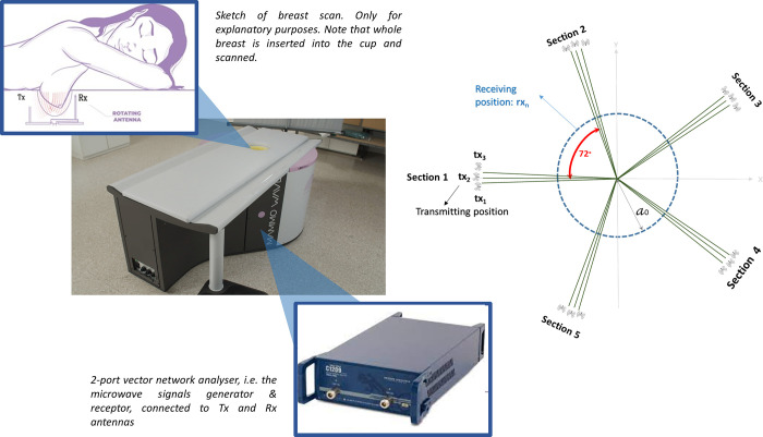
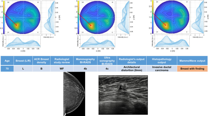
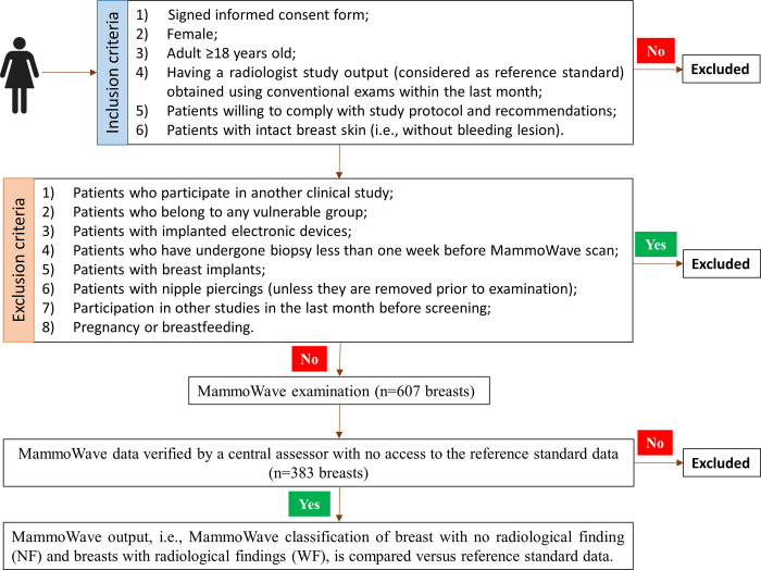
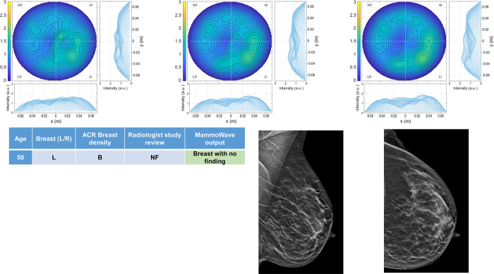
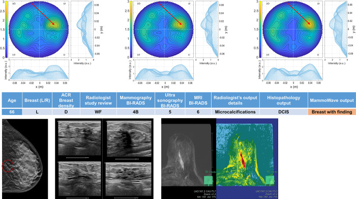
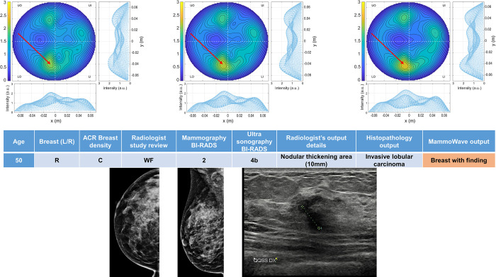
Microwave imaging is a safe and promising new technology in breast radiology, avoiding discomfort of breast compression and usage of ionizing radiation. This paper presents the first prospective microwave breast imaging study during which both symptomatic and asymptomatic subjects were recruited. Specifically, a prospective multicentre international clinical trial was performed in 2020-2021, to investigate the capability of a microwave imaging device (MammoWave) in allowing distinction between breasts with no radiological finding (NF) and breasts with radiological findings (WF), i.e., with benign or malignant lesions. Each breast scan was performed with the volunteers lying on a dedicated examination table in a comfortable prone position. MammoWave output was compared to reference standard (i.e., radiologic study obtained within the last month and integrated with histological one if available and deemed necessary by responsible investigator) to classify breasts into NF/WF categories. MammoWave output consists of a selection of microwave images' features (determined prior to trials' start), which allow distinction between NF and WF breasts (using statistical significance p<0.05). 353 women were enrolled in the study (mean age 51 years ± 12 [SD], minimum age 19, maximum age 78); MammoWave data from the first 15 women of each site, all with NF breasts, were used for calibration. Following central assessor evaluation, 111 NF (48 dense) and 272 WF (136 dense) breasts were used for comparison with MammoWave output. 272 WF comprised 182 benign findings and 90 malignant histology-confirmed cancer. A sensitivity of 82.3% was achieved (95%CI: 0.78-0.87); sensitivity is maintained when limiting the investigation to histology-confirmed breasts cancer only (90 histology-confirmed breasts cancer have been included in this analysis, having sizes ranging from 3 mm to 60 mm). Specificity value of approximately 50% was achieved as expected, since thresholds were calculated (for each feature) using median value obtained after recruiting the first 15 women (of each site), all NF. This prospective trial may represent another step for introducing microwave imaging into clinical practice, for helping in breast lesion identification in asymptomatic women.
微波成像在乳腺放射学中是一项安全且有前景的新技术,它避免了乳房压迫带来的不适以及电离辐射的使用。本文介绍了第一项前瞻性微波乳腺成像研究,该研究招募了有症状和无症状的受试者。具体而言,在2020年至2021年进行了一项前瞻性多中心国际临床试验,以研究一种微波成像设备(MammoWave)区分无放射学发现(NF)的乳房和有放射学发现(WF)的乳房的能力,即区分有良性或恶性病变的乳房。每次乳房扫描时,志愿者躺在专用检查台上,呈舒适的俯卧位。将MammoWave的输出结果与参考标准(即过去一个月内获得的放射学检查结果,并在负责的研究者认为有必要时结合组织学检查结果)进行比较,以将乳房分类为NF/WF类别。MammoWave的输出结果包括一系列微波图像特征(在试验开始前确定),这些特征能够区分NF和WF乳房(使用统计学显著性p<0.05)。353名女性参与了该研究(平均年龄51岁±12[标准差],最小年龄19岁,最大年龄78岁);每个站点的前15名女性(均为NF乳房)的数据用于校准MammoWave。经过中心评估者评估后,111个NF(48个致密型)乳房和272个WF(136个致密型)乳房用于与MammoWave的输出结果进行比较。272个WF乳房包括182个良性发现和90个经组织学证实的恶性癌症。灵敏度达到了82.3%(95%置信区间:0.78 - 0.87);当仅将研究限于组织学证实的乳腺癌时,灵敏度保持不变(本分析纳入了90个组织学证实的乳腺癌,大小范围从3毫米到60毫米)。正如预期的那样,特异性值约为50%,因为(针对每个特征)阈值是使用每个站点前15名女性(均为NF)获得的中位数计算得出的。这项前瞻性试验可能代表了将微波成像引入临床实践的又一步,有助于在无症状女性中识别乳腺病变。