Fan Zhenliang, Yang Qiaorui, Xia Hong, Zhang Peipei, Sun Ke, Yang Mengfan, Yin Riping, Zhao Dongxue, Ma Hongzhen, Shen Yiwei, Fan Junfen
Nephrology Department, The First Affiliated Hospital of Zhejiang Chinese Medical University (Zhejiang Provincial Hospital of Traditional Chinese Medicine), Hangzhou, China.
Academy of Chinese Medical Science, Zhejiang Chinese Medical University, Hangzhou, China.
Front Med (Lausanne). 2023 Jul 3;10:1066125. doi: 10.3389/fmed.2023.1066125. eCollection 2023.
Hyperplasia of the mesangial area is common in IgA nephropathy (IgAN) and diabetic nephropathy (DN), and it is often difficult to distinguish them by light microscopy alone, especially in the absence of clinical data. At present, artificial intelligence (AI) is widely used in pathological diagnosis, but mainly in tumor pathology. The application of AI in renal pathological is still in its infancy.
Patients diagnosed as IgAN or DN by renal biopsy in First Affiliated Hospital of Zhejiang Chinese Medicine University from September 1, 2020 to April 30, 2022 were selected as the training set, and patients who diagnosed from May 1, 2022 to June 30, 2022 were selected as the test set. We focused on the glomerulus and captured the field of the glomerulus in Masson staining WSI at 200x magnification, all in 1,000 × 1,000 pixels JPEG format. We augmented the data from training set through minor affine transformation, and then randomly split the training set into training and adjustment data according to 8:2. The training data and the Yolov5 6.1 algorithm were used to train the AI model with constant adjustment of parameters according to the adjusted data. Finally, we obtained the optimal model, tested this model with test set and compared it with renal pathologists.
AI can accurately detect the glomeruli. The overall accuracy of AI glomerulus detection was 98.67% and the omission rate was only 1.30%. No Intact glomerulus was missed. The overall accuracy of AI reached 73.24%, among which the accuracy of IgAN reached 77.27% and DN reached 69.59%. The AUC of IgAN was 0.733 and that of DN was 0.627. In addition, compared with renal pathologists, AI can distinguish IgAN from DN more quickly and accurately, and has higher consistency.
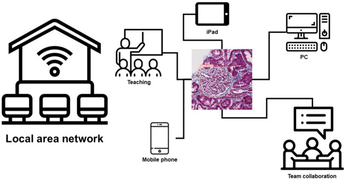
We constructed an AI model based on Masson staining images of renal tissue to distinguish IgAN from DN. This model has also been successfully deployed in the work of renal pathologists to assist them in their daily diagnosis and teaching work.
系膜区增生在IgA肾病(IgAN)和糖尿病肾病(DN)中较为常见,仅通过光学显微镜往往难以区分,尤其是在缺乏临床数据的情况下。目前,人工智能(AI)在病理诊断中广泛应用,但主要用于肿瘤病理学。AI在肾脏病理学中的应用仍处于起步阶段。
选取2020年9月1日至2022年4月30日在浙江中医药大学附属第一医院经肾活检诊断为IgAN或DN的患者作为训练集,选取2022年5月1日至2022年6月30日诊断的患者作为测试集。我们聚焦于肾小球,在200倍放大倍数下采集Masson染色全切片图像(WSI)中的肾小球视野,均为1000×1000像素的JPEG格式。我们通过轻微仿射变换对训练集数据进行增强,然后按照8:2的比例将训练集随机分为训练数据和调整数据。使用训练数据和Yolov5 6.1算法训练AI模型,并根据调整后的数据不断调整参数。最后,我们获得了最优模型,用测试集对该模型进行测试,并与肾脏病理学家进行比较。
AI能够准确检测肾小球。AI肾小球检测的总体准确率为98.67%,漏检率仅为1.30%。没有完整的肾小球被漏检。AI的总体准确率达到73.24%,其中IgAN的准确率达到77.27%,DN的准确率达到69.59%。IgAN的曲线下面积(AUC)为0.733,DN的AUC为0.627。此外,与肾脏病理学家相比,AI能够更快、更准确地区分IgAN和DN,并且具有更高的一致性。
我们构建了一个基于肾脏组织Masson染色图像的AI模型来区分IgAN和DN。该模型也已成功应用于肾脏病理学家的工作中,以协助他们的日常诊断和教学工作。