Irmandade da Santa Casa de Misericórdia de São Paulo, São Paulo, Brazil.
Hospital das Clínicas da Faculdade de Medicina da Universidade de São Paulo, São Paulo, Brazil.
BMC Musculoskelet Disord. 2023 Jul 20;24(1):596. doi: 10.1186/s12891-023-06732-z.
This study aims to evaluate the possibility of characterizing an extra-articular thickening in the knee anteromedial quadrant in routine MRI scans.
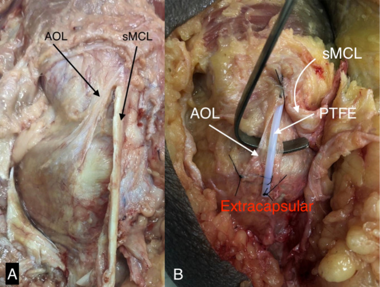
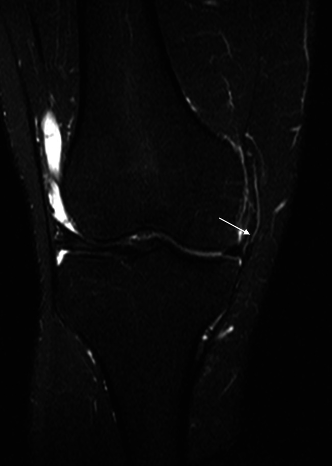
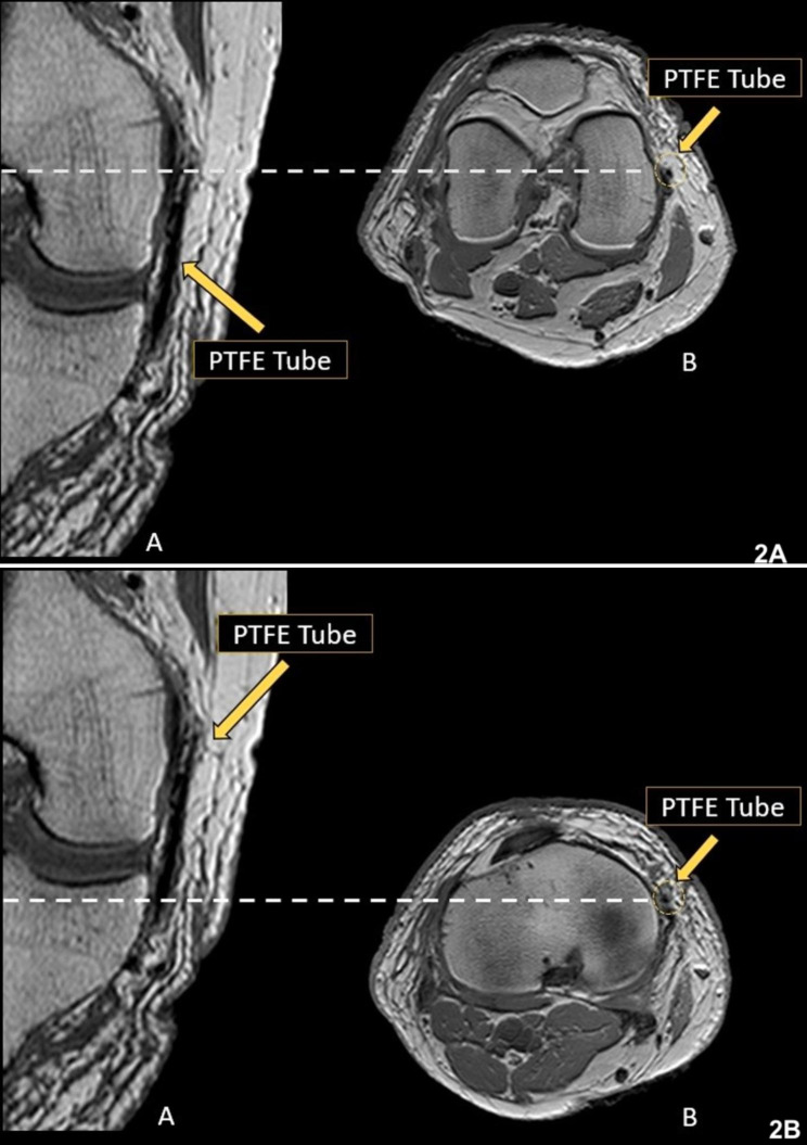
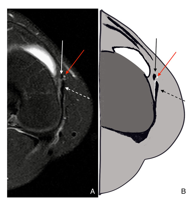
Firstly, in a pilot study, for a better understanding of this extra-articular thickening trajectory in MRI, polytetrafluoroethylene (PTFE) tubes were attached to the ligament structure topography in two dissected pieces. Afterward, 100 knee MRI studies were randomly selected from our database, and 97 met the inclusion criteria. Two musculoskeletal radiologists interpreted the exams separately. Both had previously studied the ligament in the cadaveric knee MRI with the PTFE tube.
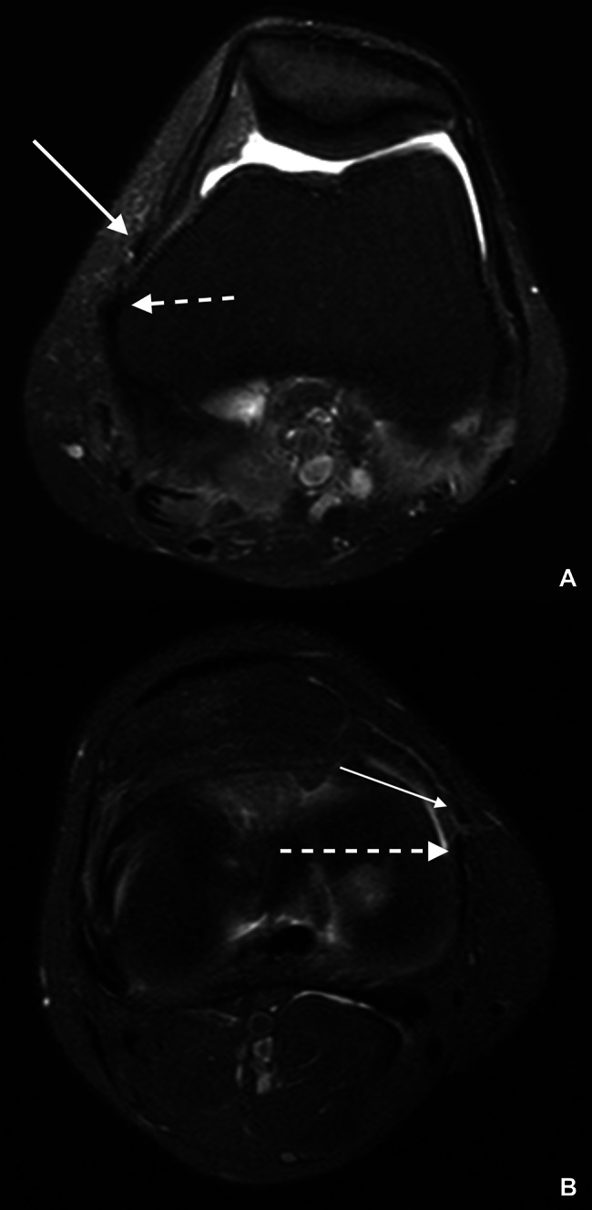
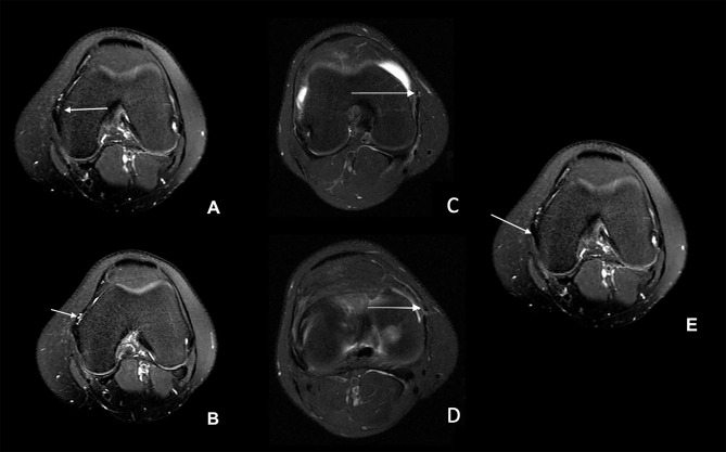
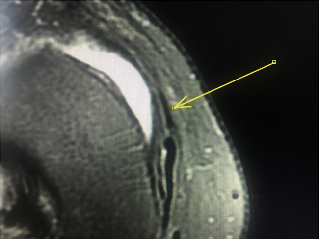
The intraobserver and interobserver agreement for the ligament identification was calculated using Cohen's Kappa coefficient. The first radiologist identified the structure in 41 of the 97 scans (42.2%), and the second radiologist in 38 scans (39.2%). The interobserver agreement was substantial, with a Kappa of 0.68 and an agreement of 84.5%. The results suggest that this extra-articular thickening, recently called Anterior Oblique Ligament (AOL) in the literature, is a structure that can be frequently visualized on MRI scans with a high level of interobserver agreement in a relatively large number of exams.
Therefore, this study indicates that MRI is a promising method for evaluating this anteromedial thickening, and it may be used for future studies of the Anterior Oblique Ligament.
本研究旨在评估在常规 MRI 扫描中对膝关节前内侧象限关节外增厚进行特征描述的可能性。
首先,在一项初步研究中,为了更好地理解 MRI 中这种关节外增厚的轨迹,将聚四氟乙烯(PTFE)管附着在两个解剖部位的韧带结构表面上。随后,从我们的数据库中随机选择了 100 例膝关节 MRI 研究,其中 97 例符合纳入标准。两名肌肉骨骼放射科医生分别对这些检查进行了解读。两位医生之前都曾在尸检膝关节 MRI 中使用 PTFE 管研究过该韧带。
使用 Cohen's Kappa 系数计算了两位观察者对韧带识别的观察者内和观察者间一致性。第一位放射科医生在 97 次扫描中的 41 次(42.2%)识别出了该结构,第二位放射科医生在 38 次扫描中(39.2%)识别出了该结构。观察者间的一致性很高,Kappa 值为 0.68,一致性为 84.5%。结果表明,这种关节外增厚,最近在文献中被称为前斜韧带(AOL),是一种在相对大量的检查中可以经常在 MRI 扫描中看到的结构,且具有很高的观察者间一致性。
因此,本研究表明 MRI 是一种评估这种前内侧增厚的有前途的方法,并且它可能用于未来对前斜韧带的研究。