Department of Otorhinolaryngology, Qilu Hospital of Shandong University, NHC Key Laboratory of Otorhinolaryngology (Shandong University), Jinan, Shandong, China.
Front Immunol. 2023 Jul 12;14:1168191. doi: 10.3389/fimmu.2023.1168191. eCollection 2023.
Human hypopharygeal squamous cell carcinoma (HSCC) is a common head and neck cancer with a poor prognosis in advanced stages. The occurrence and development of tumor is the result of mutual influence and co-evolution between tumor cells and tumor microenvironment (TME). Tumor immune microenvironment (TIME) refers to the immune microenvironment surrounding tumor cells. Studying TIME in HSCC could provide new targets and therapeutic strategies for HSCC.
We performed single-cell RNA sequencing (scRNA-seq) and analysis of hypopharyngeal carcinoma, paracancerous, and lymphoid tissues from five HSCC patients. Subdivide of B cells, T cells, macrophages cells, and monocytes and their distribution in three kinds of tissues as well as marker genes were analyzed. Different genes of IGHG1 plasma cells and SPP1+ macrophages between HSCC tissues, adjacent normal tissues and lymphatic tissues were analyzed. Additionally, we studied proliferating lymphocytes, T cells exhaustion, and T cell receptor (TCR) repertoire in three kinds of tissues.
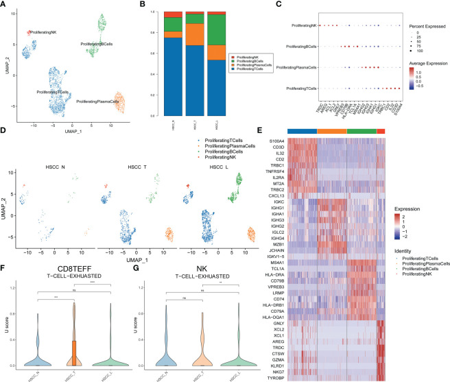
Transcriptome profiles of 132,869 single cells were obtained and grouped into seven cell clusters, including epithelial cells, lymphocytes, mononuclear phagocytics system (MPs), fibroblasts, endothelial cells (ECs), plasmacytoid dendritic cells (pDCs), and mast cells. Tumor metastasis occurred in three lymphoid tissues. Four distinct populations were identified from lymphocytes, including B cells, plasma cells, T cells and proliferating lymphocytes. We found IGHA1 and IGHG1 specific plasma cells significantly overexpressed in HSCC tissues compared with normal hypopharygeal tissues and lymphatic tissues. Five distinct populations from MPs were identified, including macrophages, monocytes, mature dendritic cells (DCs), Type 1 conventional dendritic cells (cDC1) and Type 2 conventional dendritic cells (cDC2). SPP1+ macrophages were significantly overexpressed in HSCC tissues and lymphatic tissues compared with normal hypopharygeal tissues, which are thought to be M2-type macrophages. Exhaustion of CD8+ Teff cells occurred in HSCC tissues. At last, we verified that IgA and IgG1 protein expression levels were significantly up-regulated in HSCC tissues compared to adjacent normal tissues.
Overall, this study revealed TIME in HSCC and lymphatic metastasis, and provided potential therapeutic targets for HSCC.
人类下咽鳞状细胞癌(HSCC)是一种常见的头颈部癌症,晚期预后较差。肿瘤的发生和发展是肿瘤细胞与肿瘤微环境(TME)相互影响和共同进化的结果。肿瘤免疫微环境(TIME)是指围绕肿瘤细胞的免疫微环境。研究 HSCC 的 TIME 可为 HSCC 提供新的治疗靶点和治疗策略。
我们对 5 例 HSCC 患者的下咽癌、癌旁和淋巴组织进行了单细胞 RNA 测序(scRNA-seq)和分析。分析了 B 细胞、T 细胞、巨噬细胞和单核细胞亚群及其在三种组织中的分布以及标记基因。分析了 HSCC 组织、相邻正常组织和淋巴组织中不同基因的 IGHA1 浆细胞和 SPP1+巨噬细胞。此外,我们还研究了三种组织中增殖的淋巴细胞、T 细胞耗竭和 T 细胞受体(TCR)库。
获得了 132869 个单细胞的转录组谱,并分为七个细胞群,包括上皮细胞、淋巴细胞、单核吞噬细胞系统(MPs)、成纤维细胞、内皮细胞(ECs)、浆细胞样树突状细胞(pDCs)和肥大细胞。肿瘤转移发生在三种淋巴组织中。从淋巴细胞中鉴定出四个不同的群体,包括 B 细胞、浆细胞、T 细胞和增殖淋巴细胞。我们发现与正常下咽组织和淋巴组织相比,HSCC 组织中明显过表达了 IGHA1 和 IGHG1 特异性浆细胞。从 MPs 中鉴定出五个不同的群体,包括巨噬细胞、单核细胞、成熟树突状细胞(DCs)、1 型常规树突状细胞(cDC1)和 2 型常规树突状细胞(cDC2)。与正常下咽组织相比,HSCC 组织和淋巴组织中 SPP1+巨噬细胞明显过表达,认为是 M2 型巨噬细胞。HSCC 组织中 CD8+Teff 细胞耗竭。最后,我们验证了与相邻正常组织相比,HSCC 组织中 IgA 和 IgG1 蛋白表达水平明显上调。
总之,本研究揭示了 HSCC 中的 TIME 和淋巴转移,并为 HSCC 提供了潜在的治疗靶点。