• 文献检索
  • 文档翻译
  • 深度研究
  • 学术资讯
  • Suppr Zotero 插件Zotero 插件
  • 邀请有礼
  • 套餐&价格
  • 历史记录
应用&插件
Suppr Zotero 插件Zotero 插件浏览器插件Mac 客户端Windows 客户端微信小程序
定价
高级版会员购买积分包购买API积分包
服务
文献检索文档翻译深度研究API 文档MCP 服务
关于我们
关于 Suppr公司介绍联系我们用户协议隐私条款
关注我们

Suppr 超能文献

核心技术专利:CN118964589B侵权必究
粤ICP备2023148730 号-1Suppr @ 2026

文献检索

告别复杂PubMed语法,用中文像聊天一样搜索,搜遍4000万医学文献。AI智能推荐,让科研检索更轻松。

立即免费搜索

文件翻译

保留排版,准确专业,支持PDF/Word/PPT等文件格式,支持 12+语言互译。

免费翻译文档

深度研究

AI帮你快速写综述,25分钟生成高质量综述,智能提取关键信息,辅助科研写作。

立即免费体验

基于多糖的水凝胶支架内MC3T3E1前成骨细胞球体的灌注:生物反应器尺度的实验与数值研究

Perfusion of MC3T3E1 Preosteoblast Spheroids within Polysaccharide-Based Hydrogel Scaffolds: An Experimental and Numerical Study at the Bioreactor Scale.

作者信息

Grenier Jérôme, David Bertrand, Journé Clément, Cicha Iwona, Letourneur Didier, Duval Hervé

机构信息

Laboratoire de Génie des Procédés et Matériaux, CentraleSupélec, Université Paris-Saclay, 91190 Gif-sur-Yvette, France.

Laboratoire de Mécanique Paris-Saclay, CNRS, CentraleSupélec, ENS Paris-Saclay, Université Paris-Saclay, 91190 Gif-sur-Yvette, France.

出版信息

Bioengineering (Basel). 2023 Jul 18;10(7):849. doi: 10.3390/bioengineering10070849.

DOI:10.3390/bioengineering10070849
PMID:37508876
原文链接:https://pmc.ncbi.nlm.nih.gov/articles/PMC10376891/
Abstract

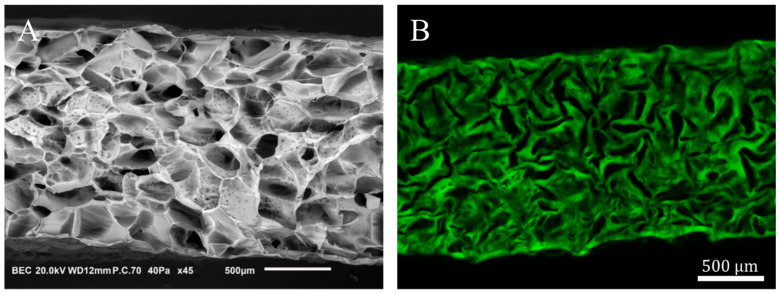
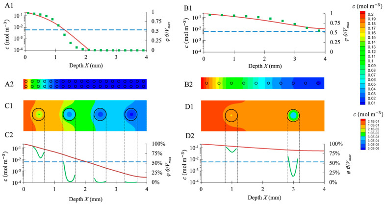
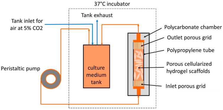
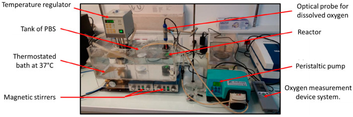
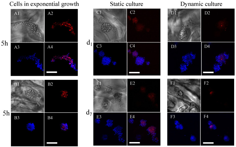
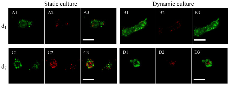
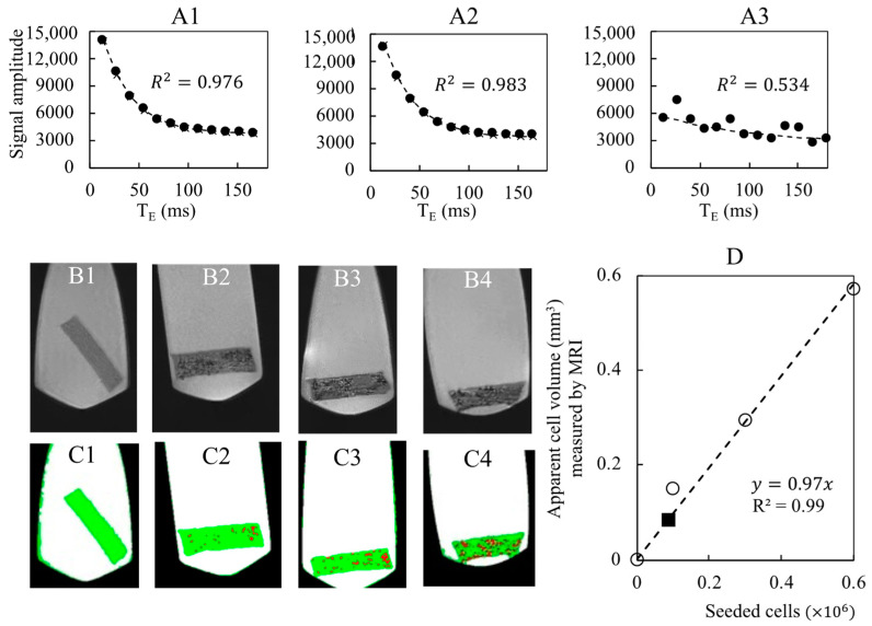
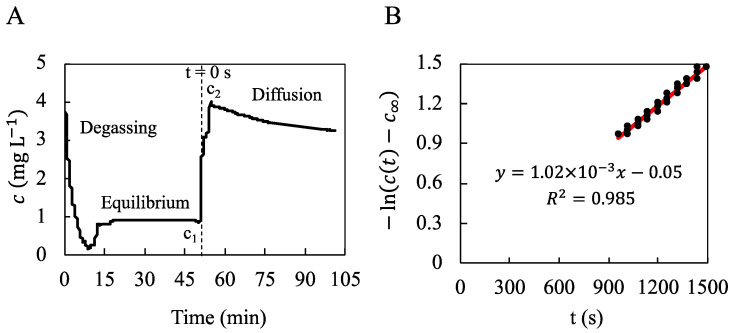
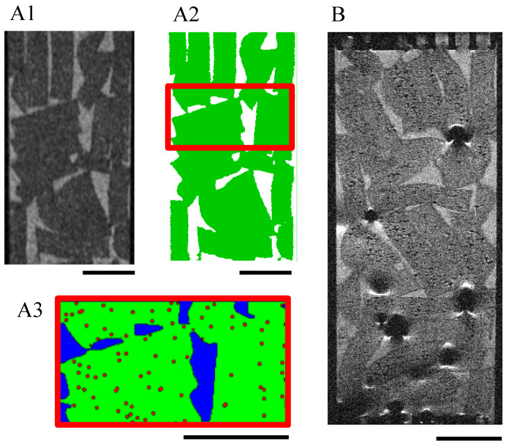
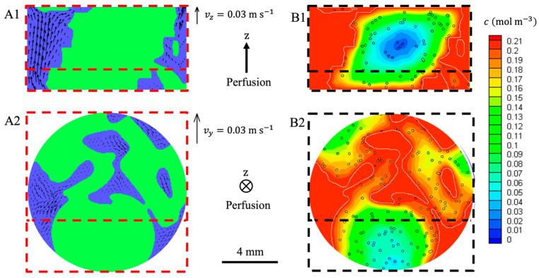
The traditional 3D culture systems in vitro lack the biological and mechanical spatiotemporal stimuli characteristic to native tissue development. In our study, we combined porous polysaccharide-based hydrogel scaffolds with a bioreactor-type perfusion device that generates favorable mechanical stresses while enhancing nutrient transfers. MC3T3E1 mouse osteoblasts were seeded in the scaffolds and cultivated for 3 weeks under dynamic conditions at a perfusion rate of 10 mL min. The spatial distribution of the cells labeled with superparamagnetic iron oxide nanoparticles was visualized by MRI. Confocal microscopy was used to assess cell numbers, their distribution inside the scaffolds, cell viability, and proliferation. The oxygen diffusion coefficient in the hydrogel was measured experimentally. Numerical simulations of the flow and oxygen transport within the bioreactor were performed using a lattice Boltzmann method with a two-relaxation time scheme. Last, the influence of cell density and spheroid size on cell oxygenation was investigated. The cells spontaneously organized into spheroids with a diameter of 30-100 μm. Cell viability remained unchanged under dynamic conditions but decreased under static culture. The cell proliferation (Ki67 expression) in spheroids was not observed. The flow simulation showed that the local fluid velocity reached 27 mm s at the height where the cross-sectional area of the flow was the smallest. The shear stress exerted by the fluid on the scaffolds may locally rise to 100 mPa, compared with the average value of 25 mPa. The oxygen diffusion coefficient in the hydrogel was 1.6×10⁻⁹ m s. The simulation of oxygen transport and consumption confirmed that the cells in spheroids did not suffer from hypoxia when the bioreactor was perfused at 10 mL min, and suggested the existence of optimal spheroid size and spacing for appropriate oxygenation. Collectively, these findings enabled us to define the optimal conditions inside the bioreactor for an efficient in vitro cell organization and survival in spheroids, which are paramount to future applications with organoids.

摘要

传统的体外3D培养系统缺乏天然组织发育所特有的生物和机械时空刺激。在我们的研究中,我们将基于多孔多糖的水凝胶支架与生物反应器型灌注装置相结合,该装置在增强营养物质传递的同时产生有利的机械应力。将MC3T3E1小鼠成骨细胞接种到支架中,并在动态条件下以10 mL/min的灌注速率培养3周。通过MRI可视化超顺磁性氧化铁纳米颗粒标记细胞的空间分布。共聚焦显微镜用于评估细胞数量、它们在支架内的分布、细胞活力和增殖情况。通过实验测量水凝胶中的氧扩散系数。使用具有双弛豫时间格式的格子玻尔兹曼方法对生物反应器内的流动和氧传输进行了数值模拟。最后,研究了细胞密度和球体大小对细胞氧合的影响。细胞自发组织成直径为30-100μm的球体。细胞活力在动态条件下保持不变,但在静态培养下降低。未观察到球体中的细胞增殖(Ki67表达)。流动模拟表明,在流动横截面积最小的高度处,局部流体速度达到27 mm/s。与25 mPa的平均值相比,流体对支架施加的剪切应力可能局部升至100 mPa。水凝胶中的氧扩散系数为1.6×10⁻⁹ m²/s。氧传输和消耗的模拟证实,当生物反应器以10 mL/min的速率灌注时,球体中的细胞不会缺氧,并表明存在最佳的球体大小和间距以实现适当的氧合。总的来说,这些发现使我们能够确定生物反应器内的最佳条件,以实现体外细胞的有效组织和球体中的存活,这对于类器官的未来应用至关重要。

https://cdn.ncbi.nlm.nih.gov/pmc/blobs/2195/10376891/6e83a8dea1fc/bioengineering-10-00849-g011.jpg
https://cdn.ncbi.nlm.nih.gov/pmc/blobs/2195/10376891/b74db05164a6/bioengineering-10-00849-g001.jpg
https://cdn.ncbi.nlm.nih.gov/pmc/blobs/2195/10376891/261f7d0a9fb6/bioengineering-10-00849-g002.jpg
https://cdn.ncbi.nlm.nih.gov/pmc/blobs/2195/10376891/bd9fc82f3931/bioengineering-10-00849-g003.jpg
https://cdn.ncbi.nlm.nih.gov/pmc/blobs/2195/10376891/81771101de80/bioengineering-10-00849-g004.jpg
https://cdn.ncbi.nlm.nih.gov/pmc/blobs/2195/10376891/d5a872cfb296/bioengineering-10-00849-g005.jpg
https://cdn.ncbi.nlm.nih.gov/pmc/blobs/2195/10376891/d39a1a4030a6/bioengineering-10-00849-g006.jpg
https://cdn.ncbi.nlm.nih.gov/pmc/blobs/2195/10376891/1b39b80764ef/bioengineering-10-00849-g007.jpg
https://cdn.ncbi.nlm.nih.gov/pmc/blobs/2195/10376891/fe917c6a702b/bioengineering-10-00849-g008.jpg
https://cdn.ncbi.nlm.nih.gov/pmc/blobs/2195/10376891/964f23c70b8f/bioengineering-10-00849-g009.jpg
https://cdn.ncbi.nlm.nih.gov/pmc/blobs/2195/10376891/6e0f4b9215af/bioengineering-10-00849-g010.jpg
https://cdn.ncbi.nlm.nih.gov/pmc/blobs/2195/10376891/6e83a8dea1fc/bioengineering-10-00849-g011.jpg

相似文献

1
Perfusion of MC3T3E1 Preosteoblast Spheroids within Polysaccharide-Based Hydrogel Scaffolds: An Experimental and Numerical Study at the Bioreactor Scale.基于多糖的水凝胶支架内MC3T3E1前成骨细胞球体的灌注:生物反应器尺度的实验与数值研究
Bioengineering (Basel). 2023 Jul 18;10(7):849. doi: 10.3390/bioengineering10070849.
2
Inlet flow rate of perfusion bioreactors affects fluid flow dynamics, but not oxygen concentration in 3D-printed scaffolds for bone tissue engineering: Computational analysis and experimental validation.灌流生物反应器入口流速影响流体动力学,但不影响 3D 打印支架中骨组织工程的氧浓度:计算分析和实验验证。
Comput Biol Med. 2020 Sep;124:103826. doi: 10.1016/j.compbiomed.2020.103826. Epub 2020 Aug 4.
3
Numerical Investigations of Hepatic Spheroids Metabolic Reactions in a Perfusion Bioreactor.灌注生物反应器中肝球体代谢反应的数值研究
Front Bioeng Biotechnol. 2019 Sep 12;7:221. doi: 10.3389/fbioe.2019.00221. eCollection 2019.
4
3-D computational modeling of media flow through scaffolds in a perfusion bioreactor.灌注生物反应器中介质流经支架的三维计算建模。
J Biomech. 2005 Mar;38(3):543-9. doi: 10.1016/j.jbiomech.2004.04.011.
5
The Hollow Porous Sphere Cell Carrier for the Dynamic Three-Dimensional Cell Culture.中空多孔球细胞载体用于动态三维细胞培养。
Tissue Eng Part C Methods. 2022 Nov;28(11):610-622. doi: 10.1089/ten.TEC.2022.0137.
6
A mathematical model and computational framework for three-dimensional chondrocyte cell growth in a porous tissue scaffold placed inside a bi-directional flow perfusion bioreactor.用于置于双向流动灌注生物反应器内的多孔组织支架中三维软骨细胞生长的数学模型和计算框架。
Biotechnol Bioeng. 2015 Dec;112(12):2601-10. doi: 10.1002/bit.25678. Epub 2015 Jul 14.
7
[Fabrication of scaffold with controlled porous structure and flow perfusion culture in vitro].[具有可控多孔结构的支架制备及体外流动灌注培养]
Sheng Wu Gong Cheng Xue Bao. 2005 Jul;21(4):579-83.
8
Numerical Simulation of Mass Transfer and Three-Dimensional Fabrication of Tissue-Engineered Cartilages Based on Chitosan/Gelatin Hybrid Hydrogel Scaffold in a Rotating Bioreactor.基于壳聚糖/明胶混合水凝胶支架在旋转生物反应器中组织工程软骨的传质数值模拟及三维制造
Appl Biochem Biotechnol. 2017 Jan;181(1):250-266. doi: 10.1007/s12010-016-2210-9. Epub 2016 Aug 15.
9
Assessment of the interplay between scaffold geometry, induced shear stresses, and cell proliferation within a packed bed perfusion bioreactor.评估在填充床灌注生物反应器中支架几何形状、诱导剪切应力和细胞增殖之间的相互作用。
Biotechnol Prog. 2019 Nov;35(6):e2880. doi: 10.1002/btpr.2880. Epub 2019 Jul 16.
10
Engineered bone culture in a perfusion bioreactor: a 2D computational study of stationary mass and momentum transport.灌注生物反应器中的工程骨培养:稳态质量和动量传递的二维计算研究。
Comput Methods Biomech Biomed Engin. 2007 Dec;10(6):429-38. doi: 10.1080/10255840701494635. Epub 2007 Aug 22.

引用本文的文献

1
Biomimetic Design and Assessment via Microenvironmental Testing: From Food Packaging Biomaterials to Implantable Medical Devices.通过微环境测试的仿生设计与评估:从食品包装生物材料到可植入医疗设备
Biomimetics (Basel). 2025 Jun 5;10(6):370. doi: 10.3390/biomimetics10060370.
2
Cancer-on-a-chip for precision cancer medicine.用于精准癌症医学的芯片上的癌症模型
Lab Chip. 2025 May 16. doi: 10.1039/d4lc01043d.
3
In vitro and in vivo assessment of a non-animal sourced chitosan scaffold loaded with xeno-free umbilical cord mesenchymal stromal cells cultured under macromolecular crowding conditions.

本文引用的文献

1
Interplay between crosslinking and ice nucleation controls the porous structure of freeze-dried hydrogel scaffolds.交联与成冰核作用之间的相互作用控制着冻干水凝胶支架的多孔结构。
Biomater Adv. 2022 Aug;139:212973. doi: 10.1016/j.bioadv.2022.212973. Epub 2022 Jun 6.
2
Engineering organoids.工程化类器官
Nat Rev Mater. 2021;6(5):402-420. doi: 10.1038/s41578-021-00279-y. Epub 2021 Feb 19.
3
Dynamic multiscale metabolic network modeling of Chinese hamster ovary cell metabolism integrating N-linked glycosylation in industrial biopharmaceutical manufacturing.
在大分子拥挤条件下培养的、负载无异种脐带间充质基质细胞的非动物源壳聚糖支架的体外和体内评估。
Biomater Biosyst. 2024 Oct 10;16:100102. doi: 10.1016/j.bbiosy.2024.100102. eCollection 2024 Dec.
4
Bone Spheroid Development Under Flow Conditions with Mesenchymal Stem Cells and Human Umbilical Vein Endothelial Cells in a 3D Porous Hydrogel Supplemented with Hydroxyapatite.在添加羟基磷灰石的三维多孔水凝胶中,间充质干细胞和人脐静脉内皮细胞在流动条件下的骨球体发育。
Gels. 2024 Oct 18;10(10):666. doi: 10.3390/gels10100666.
5
Development of Novel Polysaccharide Membranes for Guided Bone Regeneration: In Vitro and In Vivo Evaluations.用于引导骨再生的新型多糖膜的研发:体外和体内评估
Bioengineering (Basel). 2023 Oct 28;10(11):1257. doi: 10.3390/bioengineering10111257.
整合N-糖基化的中国仓鼠卵巢细胞代谢动态多尺度代谢网络建模在工业生物制药制造中的应用
Biotechnol Bioeng. 2021 Jan;118(1):397-411. doi: 10.1002/bit.27578. Epub 2020 Oct 11.
4
Computational Modeling of Human Mesenchymal Stromal Cell Proliferation and Extra-Cellular Matrix Production in 3D Porous Scaffolds in a Perfusion Bioreactor: The Effect of Growth Factors.灌注生物反应器中三维多孔支架内人骨髓间充质基质细胞增殖及细胞外基质产生的计算建模:生长因子的作用
Front Bioeng Biotechnol. 2020 Apr 29;8:376. doi: 10.3389/fbioe.2020.00376. eCollection 2020.
5
An Efficient Intestinal Organoid System of Direct Sorting to Evaluate Stem Cell Competition in Vitro.一种高效的肠类器官体外直接分选系统,用于评估干细胞竞争。
Sci Rep. 2019 Dec 30;9(1):20297. doi: 10.1038/s41598-019-55824-1.
6
Numerical Investigations of Hepatic Spheroids Metabolic Reactions in a Perfusion Bioreactor.灌注生物反应器中肝球体代谢反应的数值研究
Front Bioeng Biotechnol. 2019 Sep 12;7:221. doi: 10.3389/fbioe.2019.00221. eCollection 2019.
7
Assessment of the interplay between scaffold geometry, induced shear stresses, and cell proliferation within a packed bed perfusion bioreactor.评估在填充床灌注生物反应器中支架几何形状、诱导剪切应力和细胞增殖之间的相互作用。
Biotechnol Prog. 2019 Nov;35(6):e2880. doi: 10.1002/btpr.2880. Epub 2019 Jul 16.
8
Histological Method to Study the Effect of Shear Stress on Cell Proliferation and Tissue Morphology in a Bioreactor.组织学方法研究生物反应器中切应力对细胞增殖和组织形态的影响。
Tissue Eng Regen Med. 2019 Mar 21;16(3):225-235. doi: 10.1007/s13770-019-00181-3. eCollection 2019 Jun.
9
Engineering Stem Cell Self-organization to Build Better Organoids.工程化干细胞自组织构建更好的类器官
Cell Stem Cell. 2019 Jun 6;24(6):860-876. doi: 10.1016/j.stem.2019.05.005.
10
Mechanisms of pore formation in hydrogel scaffolds textured by freeze-drying.由冷冻干燥处理的水凝胶支架形成孔的机制。
Acta Biomater. 2019 Aug;94:195-203. doi: 10.1016/j.actbio.2019.05.070. Epub 2019 May 30.