• 文献检索
  • 文档翻译
  • 深度研究
  • 学术资讯
  • Suppr Zotero 插件Zotero 插件
  • 邀请有礼
  • 套餐&价格
  • 历史记录
应用&插件
Suppr Zotero 插件Zotero 插件浏览器插件Mac 客户端Windows 客户端微信小程序
定价
高级版会员购买积分包购买API积分包
服务
文献检索文档翻译深度研究API 文档MCP 服务
关于我们
关于 Suppr公司介绍联系我们用户协议隐私条款
关注我们

Suppr 超能文献

核心技术专利:CN118964589B侵权必究
粤ICP备2023148730 号-1Suppr @ 2026

文献检索

告别复杂PubMed语法,用中文像聊天一样搜索,搜遍4000万医学文献。AI智能推荐,让科研检索更轻松。

立即免费搜索

文件翻译

保留排版,准确专业,支持PDF/Word/PPT等文件格式,支持 12+语言互译。

免费翻译文档

深度研究

AI帮你快速写综述,25分钟生成高质量综述,智能提取关键信息,辅助科研写作。

立即免费体验

脾脏和椎旁肌表观扩散系数(ADC)测量的变异性:一项单中心大样本队列研究。

Variabilities in apparent diffusion coefficient (ADC) measurements of the spleen and the paraspinal muscle: A single center large cohort study.

作者信息

Chen Yukun, Yang Panpan, Fu Caixia, Bian Yun, Shao Chengwei, Ma Chao, Lu Jianping

机构信息

Department of Radiology, Changhai Hospital of Shanghai, Naval Medical University, Shanghai, 200433, China.

Application Developments, Siemens Shenzhen Magnetic Resonance Ltd., Siemens Healthineers, Shenzhen, 518057, China.

出版信息

Heliyon. 2023 Jul 12;9(7):e18166. doi: 10.1016/j.heliyon.2023.e18166. eCollection 2023 Jul.

DOI:10.1016/j.heliyon.2023.e18166
PMID:37519768
原文链接:https://pmc.ncbi.nlm.nih.gov/articles/PMC10372245/
Abstract

PURPOSE

Evaluation of the variabilities in apparent diffusion coefficient (ADC) measurements of the spleen (ADC) and the paraspinal muscles (ADC) to identify the reference organ for normalizing the ADC from the abdominal diffusion weighted imaging (DWI).

METHODS

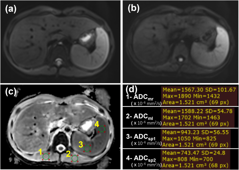
Two MRI scanners, with 314 abdominal exams on the GE and 929 on the Siemens system, were used for MRI examinations including DWI (b-values, 50 and 800 s/mm). For a subset of 73 exams on the Siemens system a second exam was conducted. Four regions of interest (ROIs) in each exam were placed to measure the ADC and the bilateral ADC. ADC variability between patients (on each scanner separately), ADC variability due to ROI placement between the two ROIs in each organ, and variability in the subset between the first and second exams were assessed.

RESULTS

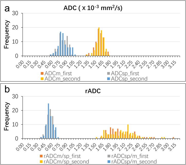
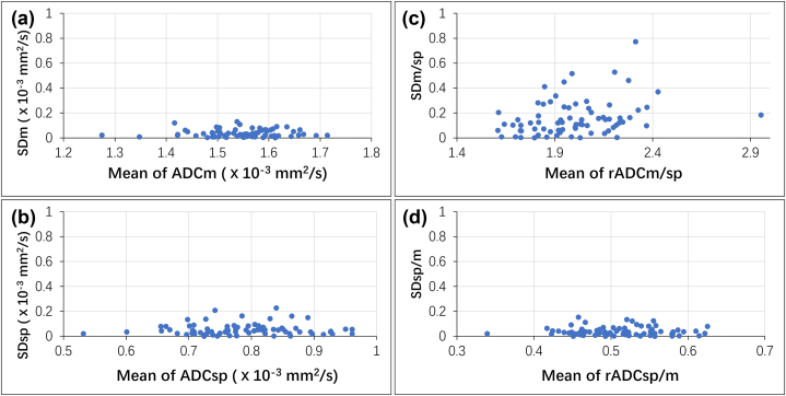
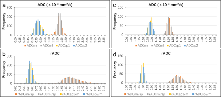
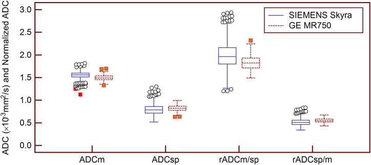
The ADC was more scattered and variable than the ADC in the comparability (n = 929 and 314 for two MRI scanners, respectively) and repeatability (n = 73) datasets. The Bland-Altmann bias and limits of agreement (LoAs) for the ADC (ICC, 0.47; CV, 0.070) and ADC (ICC, 0.67; CV, 0.023) in the repeatability datasets (n = 73) were -0.1 (-25.7%-25.6%) and -0.3 (-8.8%-8.1%), respectively. For the Siemens system, the Bland-Altmann bias and LoAs for the ADC (ICC, 0.72; CV, 0.061) and ADC (ICC, 0.53; CV, 0.030) in the comparability datasets (n = 929) were 2.1 (-20.0%-24.2%) and 0.7 (-10.0%-11.4%), respectively. Similar findings have been found in the GE system (n = 314). The CVs for the ADC measurements were lower than those of the ADC both in the repeatability and the comparability analyses (all p < 0.001).

CONCLUSION

Paraspinal muscles demonstrate better reference characteristics than the spleen in estimating ADC variability of abdominal DWI.

摘要

目的

评估脾脏表观扩散系数(ADC)测量值和椎旁肌ADC测量值的变异性,以确定用于标准化腹部扩散加权成像(DWI)中ADC的参考器官。

方法

使用两台MRI扫描仪,GE系统上进行了314例腹部检查,西门子系统上进行了929例。MRI检查包括DWI(b值为50和800 s/mm²)。在西门子系统的73例检查子集中进行了第二次检查。每次检查中放置四个感兴趣区域(ROI)来测量ADC和双侧ADC。评估了患者之间(分别在每台扫描仪上)的ADC变异性、每个器官中两个ROI之间由于ROI放置导致的ADC变异性以及第一次和第二次检查之间子集中的变异性。

结果

在可比性数据集(两台MRI扫描仪分别为n = 929和314)和重复性数据集(n = 73)中,脾脏ADC比椎旁肌ADC更分散且变异性更大。重复性数据集(n = 73)中脾脏ADC(组内相关系数[ICC],0.47;变异系数[CV],0.070)和椎旁肌ADC(ICC,0.67;CV,0.023)的Bland-Altmann偏差和一致性界限(LoA)分别为-0.1(-25.7%至25.6%)和-0.3(-8.8%至8.1%)。对于西门子系统,可比性数据集(n = 929)中脾脏ADC(ICC,0.72;CV,0.061)和椎旁肌ADC(ICC,0.53;CV,0.030)的Bland-Altmann偏差和LoA分别为2.1(-20.0%至24.2%)和0.7(-10.0%至11.4%)。GE系统(n = 314)中也发现了类似结果。在重复性和可比性分析中,脾脏ADC测量的CV均低于椎旁肌ADC的CV(所有p < 0.001)。

结论

在估计腹部DWI的ADC变异性方面,椎旁肌比脾脏表现出更好的参考特征。

https://cdn.ncbi.nlm.nih.gov/pmc/blobs/d93d/10372245/ec898bbc2887/gr7.jpg
https://cdn.ncbi.nlm.nih.gov/pmc/blobs/d93d/10372245/0723c3439927/gr1.jpg
https://cdn.ncbi.nlm.nih.gov/pmc/blobs/d93d/10372245/8529a025b4c4/gr2.jpg
https://cdn.ncbi.nlm.nih.gov/pmc/blobs/d93d/10372245/7df000e847f4/gr3.jpg
https://cdn.ncbi.nlm.nih.gov/pmc/blobs/d93d/10372245/3ce6ff6eb860/gr4.jpg
https://cdn.ncbi.nlm.nih.gov/pmc/blobs/d93d/10372245/e02a6c4ba6ec/gr5.jpg
https://cdn.ncbi.nlm.nih.gov/pmc/blobs/d93d/10372245/c74a221ef32f/gr6.jpg
https://cdn.ncbi.nlm.nih.gov/pmc/blobs/d93d/10372245/ec898bbc2887/gr7.jpg

相似文献

1
Variabilities in apparent diffusion coefficient (ADC) measurements of the spleen and the paraspinal muscle: A single center large cohort study.脾脏和椎旁肌表观扩散系数(ADC)测量的变异性:一项单中心大样本队列研究。
Heliyon. 2023 Jul 12;9(7):e18166. doi: 10.1016/j.heliyon.2023.e18166. eCollection 2023 Jul.
2
Apparent diffusion coefficient normalization of normal liver: Will it improve the reproducibility of diffusion-weighted imaging at different MR scanners as a new biomarker?正常肝脏的表观扩散系数标准化:作为一种新的生物标志物,它能否提高不同磁共振扫描仪上扩散加权成像的可重复性?
Medicine (Baltimore). 2017 Jan;96(3):e5910. doi: 10.1097/MD.0000000000005910.
3
Diffusion-weighted MR imaging of upper abdominal organs at different time points: Apparent diffusion coefficient normalization using a reference organ.不同时间点上腹部器官的扩散加权磁共振成像:使用参考器官进行表观扩散系数标准化
J Magn Reson Imaging. 2017 May;45(5):1494-1501. doi: 10.1002/jmri.25456. Epub 2016 Sep 13.
4
Repeatability of Apparent Diffusion Coefficient at 3.0 Tesla in Normal Pancreas.3.0特斯拉下正常胰腺表观扩散系数的可重复性
Cureus. 2021 Jun 18;13(6):e15734. doi: 10.7759/cureus.15734. eCollection 2021 Jun.
5
Repeatability and reproducibility of ADC measurements: a prospective multicenter whole-body-MRI study.ADC 测量的可重复性和再现性:一项前瞻性多中心全身 MRI 研究。
Eur Radiol. 2021 Jul;31(7):4514-4527. doi: 10.1007/s00330-020-07522-0. Epub 2021 Jan 6.
6
Diffusion-weighted quantitative MRI of pleural abnormalities: Intra- and interobserver variability in the apparent diffusion coefficient measurements.胸腔异常的弥散加权定量 MRI:表观弥散系数测量的观察者内和观察者间可变性。
J Magn Reson Imaging. 2017 Sep;46(3):769-782. doi: 10.1002/jmri.25633. Epub 2017 Jan 24.
7
Does positron emission tomography data acquisition impact simultaneous diffusion-weighted imaging in a whole-body PET/MRI system?正电子发射断层扫描数据采集是否会影响全身 PET/MRI 系统中的扩散加权成像?
Eur J Radiol. 2013 Feb;82(2):380-4. doi: 10.1016/j.ejrad.2012.11.018. Epub 2012 Dec 10.
8
Repeatability and Reproducibility of ADC Measurements and MRI Signal Intensity Measurements of Bone Marrow in Monoclonal Plasma Cell Disorders: A Prospective Bi-institutional Multiscanner, Multiprotocol Study.单克隆浆细胞疾病中骨髓ADC测量和MRI信号强度测量的可重复性和再现性:一项前瞻性双机构多扫描仪、多协议研究
Invest Radiol. 2022 Apr 1;57(4):272-281. doi: 10.1097/RLI.0000000000000838.
9
Preliminary Experience of 5.0 T Higher Field Abdominal Diffusion-Weighted MRI: Agreement of Apparent Diffusion Coefficient With 3.0 T Imaging.5.0T 高场腹部扩散加权 MRI 的初步经验:表观扩散系数与 3.0T 成像的一致性。
J Magn Reson Imaging. 2022 Oct;56(4):1009-1017. doi: 10.1002/jmri.28097. Epub 2022 Feb 4.
10
Intravoxel incoherent motion diffusion-weighted MR imaging of the liver: effect of triggering methods on regional variability and measurement repeatability of quantitative parameters.肝脏体素内不相干运动弥散加权磁共振成像:触发方法对定量参数的区域变异性和测量重复性的影响。
Radiology. 2015 Feb;274(2):405-15. doi: 10.1148/radiol.14140759. Epub 2014 Sep 17.

引用本文的文献

1
The relevance of T2 relaxation time in interpreting MRI apparent diffusion coefficient (ADC) map for musculoskeletal structures.T2弛豫时间在解读肌肉骨骼结构的MRI表观扩散系数(ADC)图中的相关性。
Quant Imaging Med Surg. 2023 Dec 1;13(12):7657-7666. doi: 10.21037/qims-23-1392. Epub 2023 Oct 20.

本文引用的文献

1
Technical Basics of Diffusion-Weighted Imaging.扩散加权成像的技术基础
Magn Reson Imaging Clin N Am. 2021 May;29(2):129-136. doi: 10.1016/j.mric.2021.01.001. Epub 2021 Mar 23.
2
Diffusion-weighted magnetic resonance imaging for the assessment of liver fibrosis in chronic viral hepatitis.弥散加权磁共振成像在慢性病毒性肝炎肝纤维化评估中的应用。
PLoS One. 2021 Mar 4;16(3):e0248024. doi: 10.1371/journal.pone.0248024. eCollection 2021.
3
Advanced Diffusion-Weighted Abdominal Imaging: Qualitative and Quantitative Comparison of High and Ultra-High b-Values for Lesion Detection and Image Quality.
高级扩散加权腹部成像:高和超高 b 值在病变检测和图像质量方面的定性和定量比较。
Invest Radiol. 2020 May;55(5):285-292. doi: 10.1097/RLI.0000000000000639.
4
Diffusion-weighted imaging of the breast-a consensus and mission statement from the EUSOBI International Breast Diffusion-Weighted Imaging working group.乳腺弥散加权成像的共识和任务声明:来自 EUSOBI 国际乳腺弥散加权成像工作组。
Eur Radiol. 2020 Mar;30(3):1436-1450. doi: 10.1007/s00330-019-06510-3. Epub 2019 Nov 30.
5
Consensus-based technical recommendations for clinical translation of renal diffusion-weighted MRI.基于共识的肾脏弥散加权 MRI 临床转化技术推荐
MAGMA. 2020 Feb;33(1):177-195. doi: 10.1007/s10334-019-00790-y. Epub 2019 Nov 1.
6
Reproducibility of normalized apparent diffusion coefficient measurements on 3.0-T diffusion-weighted imaging of normal pancreas in a healthy population.健康人群中3.0-T磁共振扩散加权成像对正常胰腺标准化表观扩散系数测量的可重复性
Medicine (Baltimore). 2019 Apr;98(14):e15104. doi: 10.1097/MD.0000000000015104.
7
Intra-individual comparison of conventional and simultaneous multislice-accelerated diffusion-weighted imaging in upper abdominal solid organs: value of ADC normalization using the spleen as a reference organ.上腹部实质脏器常规与单次激发多层加速扩散加权成像的个体内比较:应用脾脏作为参照器官进行 ADC 值标准化的价值。
Abdom Radiol (NY). 2019 May;44(5):1808-1815. doi: 10.1007/s00261-019-01924-5.
8
Variability in quantitative diffusion-weighted MR imaging (DWI) across different scanners and imaging sites: is there a potential consensus that can help reducing the limits of expected bias?不同扫描仪和成像部位的定量扩散加权磁共振成像(DWI)的可变性:是否存在潜在的共识,可以帮助减少预期偏差的限制?
Eur Radiol. 2019 May;29(5):2243-2245. doi: 10.1007/s00330-018-5866-4. Epub 2018 Nov 28.
9
Quantitative imaging biomarkers alliance (QIBA) recommendations for improved precision of DWI and DCE-MRI derived biomarkers in multicenter oncology trials.定量成像生物标志物联盟(QIBA)关于提高多中心肿瘤试验中 DWI 和 DCE-MRI 衍生生物标志物精度的建议。
J Magn Reson Imaging. 2019 Jun;49(7):e101-e121. doi: 10.1002/jmri.26518. Epub 2018 Nov 19.
10
Diffusion MRI of cancer: From low to high b-values.癌症的扩散 MRI:从低到高 b 值。
J Magn Reson Imaging. 2019 Jan;49(1):23-40. doi: 10.1002/jmri.26293. Epub 2018 Oct 12.