Key Laboratory of Biomedical Information Engineering of Ministry of Education, Department of Biomedical Engineering, School of Life Science and Technology, Xi'an Jiaotong University, Xi'an, China.
Department of Radiology and Biomedical Research Imaging Center, the University of North Carolina at Chapel Hill, Chapel Hill, United States.
Elife. 2023 Aug 1;12:e75401. doi: 10.7554/eLife.75401.
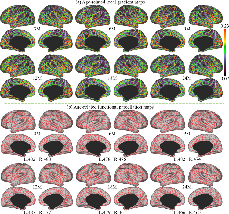
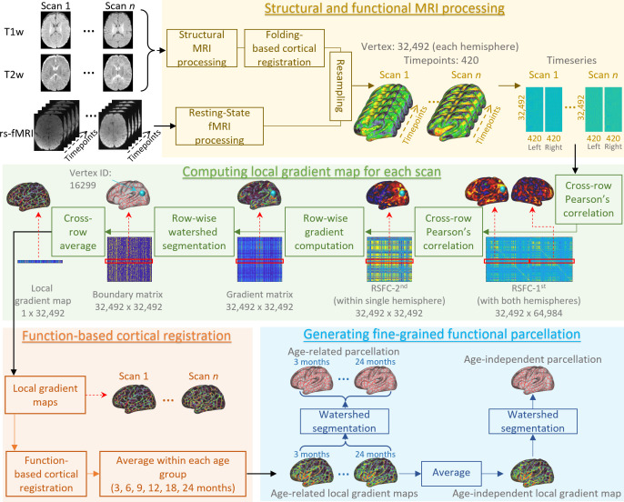
Resting-state functional MRI (rs-fMRI) is widely used to examine the dynamic brain functional development of infants, but these studies typically require precise cortical parcellation maps, which cannot be directly borrowed from adult-based functional parcellation maps due to the substantial differences in functional brain organization between infants and adults. Creating infant-specific cortical parcellation maps is thus highly desired but remains challenging due to difficulties in acquiring and processing infant brain MRIs. In this study, we leveraged 1064 high-resolution longitudinal rs-fMRIs from 197 typically developing infants and toddlers from birth to 24 months who participated in the Baby Connectome Project to develop the first set of infant-specific, fine-grained, surface-based cortical functional parcellation maps. To establish meaningful cortical functional correspondence across individuals, we performed cortical co-registration using both the cortical folding geometric features and the local gradient of functional connectivity (FC). Then we generated both age-related and age-independent cortical parcellation maps with over 800 fine-grained parcels during infancy based on aligned and averaged local gradient maps of FC across individuals. These parcellation maps reveal complex functional developmental patterns, such as changes in local gradient, network size, and local efficiency, especially during the first 9 postnatal months. Our generated fine-grained infant cortical functional parcellation maps are publicly available at https://www.nitrc.org/projects/infantsurfatlas/ for advancing the pediatric neuroimaging field.
静息态功能磁共振成像(rs-fMRI)被广泛用于研究婴儿大脑的动态功能发育,但这些研究通常需要精确的皮质分割图谱,由于婴儿和成人的大脑功能组织存在显著差异,因此不能直接从基于成人的功能分割图谱中借用。因此,创建特定于婴儿的皮质分割图谱是非常需要的,但由于获取和处理婴儿脑 MRI 的困难,这仍然是一个挑战。在这项研究中,我们利用了 197 名来自婴儿连接组计划的正常发育的婴儿和幼儿从出生到 24 个月的 1064 次高分辨率纵向 rs-fMRI,开发了第一套特定于婴儿的、细粒度的、基于表面的皮质功能分割图谱。为了在个体之间建立有意义的皮质功能对应关系,我们使用皮质折叠的几何特征和功能连接(FC)的局部梯度进行了皮质配准。然后,我们基于个体间对齐和平均的 FC 局部梯度图,在婴儿期生成了超过 800 个细粒度分区的与年龄相关和与年龄无关的皮质分区图。这些分区图揭示了复杂的功能发育模式,例如局部梯度、网络大小和局部效率的变化,特别是在出生后的前 9 个月。我们生成的精细的婴儿皮质功能分区图谱可在 https://www.nitrc.org/projects/infantsurfatlas/ 上公开获取,用于推进儿科神经影像学领域的发展。